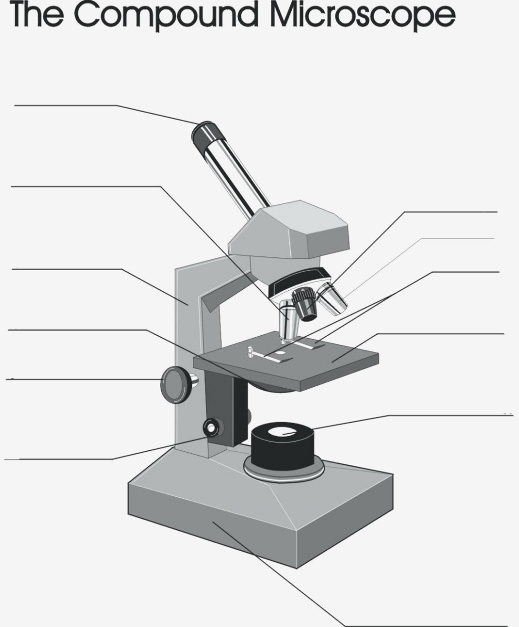
38 labelled compound microscope
Compound Microscope Parts - Labeled Diagram and their Functions - Rs ... The term "compound" refers to the microscope having more than one lens. Basically, compound microscopes generate magnified images through an aligned pair of the objective lens and the ocular lens. In contrast, "simple microscopes" have only one convex lens and function more like glass magnifiers. (a) Draw a labelled ray diagram of a compound microscope. (b) … 23.05.2018 · (a) Labelled diagram of compound microscope. The objective lens form image A' B' near the first focal point of eyepiece. (b) Angular magnification of objective lens m 0 = linear magnification h'/h. where L is the distance between second focal point of the objective and first focal point of eyepiece.If the final image A'' B'' is formed at the near point.
Draw a neat labelled diagram of a compound microscope class ... - Vedantu Physics. Grade 12. Compound Microscope. Book Online Demo. Answer. Draw a neat labelled diagram of a compound microscope. Derive the magnifying power for it. A telescope has an objective of focal length 140cm and an eyepiece of focal length 5cm. Find the magnifying power and separation between objective and eyepiece.
Labelled compound microscope
Compound Microscope Labeled : Best Quality Product [2022] Products Suggest. By, John Boyne. Jun 15, 2022. TOP Choice #1. OMAX 40X-2000X LED Binocular Compound Lab Microscope w/ Double Layer Mechanical Stage + Blank Slides, Cover Slips, & Lens Cleaning Paper, M82ES-SC100-LP100. Product Highlights. Our Score: 9.8. microscopeinternational.com › fluorescence-microscopyExplanation and Labelled Images - New York Microscope Company Dec 16, 2020 · A fluorescence microscope uses a higher intensity light to illuminate the samples. Parts of a Fluorescence Microscope A powerful light source (xenon or mercury arch lamp) : The light emitted from the mercury arc lamp is 10-100 times brighter than most incandescent lamps and provides light in a wide range of wavelengths, from ultra-violet to the ... › 59923 › draw-labelled-ray-diagram(a) Draw a labelled ray diagram of a compound microscope. (b ... May 23, 2018 · Thus, total magnification of the compound microscope. M = m 0 x m e = L/f 0 x D/f e (c) Aperture and focal length increase or decrease the resolving power of the compound microscope. Resolving power of microscope is given by. R.P. = (2n sin θ)/(1.22λ)
Labelled compound microscope. › ask › questionA compound microscope uses an objective lens of focal length ... (a) Draw the labelled ray diagram for the formation of image by a compound microscope. Derive an expression for its total magnification (or magnifying power), when the final image is formed at the near point. (b) Why both objective and eyepiece of a compound microscope must have short focal lengths? Compound Microscope Parts, Functions, and Labeled Diagram Compound Microscope Definitions for Labels. Eyepiece (ocular lens) with or without Pointer: The part that is looked through at the top of the compound microscope. Eyepieces typically have a magnification between 5x & 30x. Monocular or Binocular Head: Structural support that holds & connects the eyepieces to the objective lenses. Compound Microscope - Types, Parts, Diagram, Functions and Uses Compound microscope - It has two convex lenses. It is called a compound microscope because it compounds the light as it passes through the lenses to magnify. The image of the object being viewed is enlarged because of the lens near the object. An eyepiece, an additional lens, is where real magnification takes place. Class-X Science-086 SAMPLE QUESTION PAPER-19 TIME: 3 Hrs. 17. A compound A (C 2H 4O 2) reacts with Na metal to form a compound ‘B’ and evolves a gas which burns with a pop sound. Compound ‘A’ on treatment with an alcohol ‘C’ in presence of an acid forms a sweet smelling compound ‘D’ (C 4H 8O 2). On addition of NaOH to ‘D’ gives back B and C. Identify A, B, C and D write the ...
Compound Microscope- Definition, Labeled Diagram, Principle, Parts, Uses A compound microscope is of great use in pathology labs so as to identify diseases. Various crime cases are detected and solved by drawing out human cells and examining them under the microscope in forensic laboratories. The presence or absence of minerals and the presence of metals can be identified using compound microscopes. learn.careers360.com › school › question-a-draw-a(a) Draw a labelled ray diagram of compound microscope, when ... (a) Draw a labelled ray diagram of compound microscope, when final image forms at the least distance of distinct vision. (b) Why is its objective of short focal length and of short aperture, compared to its eyepiece? Explain. (c) The focal length of the objective is 4 cm while that of eyepiece is 10 cm. The object is placed at a distance of 6 cm from the objective lens. (i) Calculate the ... Labelled Diagram of Compound Microscope - Biology Discussion The below mentioned article provides a labelled diagram of compound microscope. Part # 1. The Stand: The stand is made up of a heavy foot which carries a curved inclinable limb or arm bearing the body tube. The foot is generally horse shoe-shaped structure (Fig. 2) which rests on table top or any other surface on which the microscope in kept. Label the microscope — Science Learning Hub All microscopes share features in common. In this interactive, you can label the different parts of a microscope. Use this with the Microscope parts activity to help students identify and label the main parts of a microscope and then describe their functions. Drag and drop the text labels onto the microscope diagram.
Parts of a Compound Microscope - Labeled (with diagrams) A compound microscope is known as a high-power microscope that enables you to achieve a high level of magnification. Smaller specimens can be thoroughly viewed using a compound microscope. ... Image 3: A compound microscope with a corresponding label of the different parts. imagesource: images.slideplayer.com. The optical components of a ... (i) Draw a neat labelled ray diagram of a compound microscope. Explain ... Working: Suppose a small object AB is placed slightly away from the first focus F 0 ' of the objective lens. The objective lens forms the real, inverted and magnified image A'B', which acts as an object for eyepiece. The eyepiece is so adjusted that the image A'B' lies between the first focus F e ' and the eyepiece E. The eyepiece forms its image A'' B'' which is virtual, erect and magnified. A compound microscope uses an objective lens of focal length 4 … Click here👆to get an answer to your question ️ A compound microscope uses an objective lens of focal length 4 cm and eyepiece lens of focal length 10 cm . An object is placed at 6 cm from the objective lens. Calculate the magnifying power of the compound microscope. Also, calculate the length of the microscope. Parts of a Compound Microscope (And their Functions) List of Microscope Parts and their Functions. 1. Ocular Tubes (Monocular, Binocular & Trinocular) The ocular tubes, are to tubes that lead from the head of the microscope out to your eyes. On the end of the ocular tubes are usually interchangeable eyepieces (commonly 10X and 20X) that increase magnification.
microscopewiki.com › compound-microscopeCompound Microscope – Diagram (Parts labelled), Principle and ... Feb 03, 2022 · Using a combination of lenses, the working principle of a compound microscope is that a highly magnified image of the specimen is formed at the least possible distance from the distinct vision of an eye that is held very close to the eyepiece of the microscope when the specimen is placed just beyond the focus of the objective lens.
How to draw compound of Microscope easily - step by step - YouTube I will show you " How to draw compound of microscope easily - step by step "Please watch carefully and try this okay.Thanks for watching.....#microscopedrawi...
Draw a neat labelled diagram of a compound microscope and explain its ... Description : It consists of two convex lenses separated by a distance. The lens near the object is called objective and the lens near the eye is called eye piece. The objective lens has small focal length and eye piece has of larger focal length. The distance of the object can be adjusted by means of a rack and pinion arrangement.
Compound Microscope – Diagram (Parts labelled), Principle and … 03.02.2022 · A compound microscope basically consists of optical and structural components. Within these two systems, there are multiple components within them and they are: Image : Labeled Diagram of compound microscope parts. See: Labeled Diagram showing differences between compound and simple microscope parts Structural Components. The three …
The Parts of a Compound Microscope and How To Handle … We shall only learn about the compound light microscope. It uses visible light to visualize the specimen, but passes that light through two separate lens to magnify the image. The compound microscopes we will use in this course are sturdy instruments but they still have a lot of moving parts. They can be damaged and broken through misuse and mishandling. A large part of …
Parts of a Compound Microscope and Their Functions Compound microscope magnification is determined by multiplying the eyepiece and objective powers. When viewed through a 5X eyepiece with a 10X objective, an item is magnified 5 x 10=50 times. The magnification is 10 x 45 = 450 times when using a 10X eyepiece and a 45X objective. How to Use the Compound Microscope
Microscope Parts, Function, & Labeled Diagram - slidingmotion Microscope parts labeled diagram gives us all the information about its parts and their position in the microscope. Microscope Parts Labeled Diagram The principle of the Microscope gives you an exact reason to use it. It works on the 3 principles. Magnification Resolving Power Numerical Aperture. Parts of Microscope Head Base Arm Eyepiece Lens
(a) Draw a labelled ray diagram of compound microscope, when … (a) Draw a labelled ray diagram of compound microscope, when final image forms at the least distance of distinct vision. (b) Why is its objective of short focal length and of short aperture, compared to its eyepiece? Explain. (c) The focal length of the objective is 4 cm while that of eyepiece is 10 cm. The object is placed at a distance of 6 cm from the objective lens. (i) …
Inverted Microscope - Advantages, Disadvantages Merely a series of lens in a tube, it was the forerunner of today’s compound microscope. Inverted Microscope Labelled Diagram from olympusmicro.com. A light source was added by Anton van Leeuwenhoek, and the lenses improved, in the late 1600s. However, until the 1800s there were few major improvements in the light microscope. Then in 1850, John Lawrence …


Post a Comment for "38 labelled compound microscope"