45 eye diagram with labelling
Labelling the eye — Science Learning Hub In this interactive, you can label parts of the human eye. Use your mouse or finger to hover over a box to highlight the part to be named. Drag and drop the text labels onto the boxes next to the eye diagram If you want to redo an answer, click on the box and the answer will go back to the top so you can move it to another box. Diagram of the Eye - Home - Lions Eye Institute Instructions. Click the parts of the eye to see a description for each. Hover the diagram to zoom. Iris. The iris is the coloured part of the eye which surrounds the pupil. It controls light levels inside the eye, similar to the aperture on a camera. The iris contains tiny muscles that widen and narrow the pupil size.
Eye Diagram Quiz - ProProfs Quiz Try this amazing Eye Diagram Quiz quiz which has been attempted 4326 times by avid quiz takers. Also explore over 77 similar quizzes in this category. Take Quizzes. Animal; Nutrition; ... Quiz: Label The Parts Of The Eye. People say that the eyes are the windows to a person's soul. In the class today, we covered parts of the eye, and what ...

Eye diagram with labelling
How To Draw A Eye And Ear And Label Them All The ears top should be along the eyebrow line and the bottom should closely line up to the nose. Parts of the Ear. Eyes drawing - step 2. Label The Parts Of The Eye. Okay shade in the inside outer edge of the inner circle of the eye yes that is a lot of words. Select the correct label for each part of the ear. Labelling the eye — Science Learning Hub Labelling the eye Add to collection The human eye contains structures that allow it to perceive light, movement and colour differences. In this activity, students use online or paper resources to identity and label the main parts of the human eye. By the end of this activity, students should be able to: identify the main parts of the human eye Structure and Functions of Human Eye with labelled Diagram Structure and Functions of Human Eye with labelled Diagram Biology Biology Article Structure Of Eye Structure of the Eye The eye is one of the sensory organs of the body. In this article, we shall explore the anatomy of the eye The structure of the eye is an important topic to understand as it one of the important sensory organs in the human body.
Eye diagram with labelling. Diagram Of Human Eye With Labelling : Diagram Human Eye Anatomy Label ... Diagram Of Human Eye With Labelling : Diagram Human Eye Anatomy Label Illustration Stock Vector Royalty Free 1888701397. Here are some tips for identifying common eye problems and how to treat them. It is a transparent, biconvex, lens of an eye. Students label and color the image for practice with anatomy. Download a free preview or high ... Eye Anatomy: 16 Parts of the Eye & Their Functions The following are parts of the human eyes and their functions: 1. Conjunctiva. The conjunctiva is the membrane covering the sclera (white portion of your eye). The conjunctiva also covers the interior of your eyelids. Conjunctivitis, often known as pink eye, occurs when this thin membrane becomes inflamed or swollen. Eye diagram basics: Reading and applying eye diagrams - EDN Interpreting an eye diagram. A properly constructed eye should contain every possible bit sequence from simple alternate 1's and 0's to isolated 1's after long runs of 0's, and all other patterns that may show up weaknesses in the design. Eye diagrams usually include voltage and time samples of the data acquired at some sample rate ... Eye Diagram - Differentiated Worksheets and EASEL Activities Use this simple eye diagram for primary students as they learn about the human eye. Two differentiated worksheets included: one with a word bank and one without. Words to label: eyebrow, eyelid, eyelashes, pupil, iris, and sclera. Find this Pin and more on Ylli by Kirsi Liuska. Eye Anatomy Diagram. Ear Diagram. Science Student.
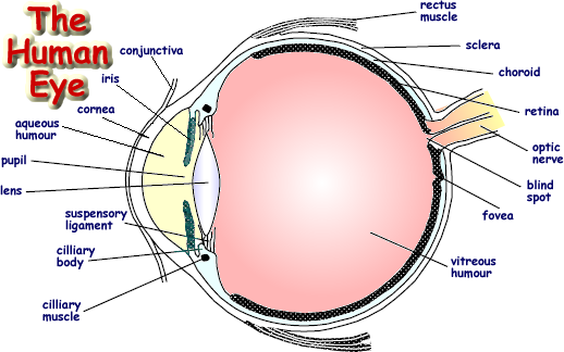
Draw And Label The Structure Of The Human Eye A human eye is roughly 23 cm in diameter and is almost a spherical ball filled with some fluid. It is the outer covering a protective tough white layer called the sclera white part of the eye. Cornea Iris Pupil Ciliary muscles Eye lens retina and optical nerves which are labelled in the diagram below. FREE! - The Human Eye Labeling Activity (Teacher-Made) In this resource, you'll find a 2-page PDF that is easy to download, print out, and use immediately with your class. The first page is a labelling exercise with two diagrams of the human eye. One is a view from the outside, and the other is a more detailed cross-section. On the second page, you'll find a set of answers showing the properly labelled human eyes, designed to help you check ... The Eye - Science Quiz - GeoGuessr The cornea is a clear covering that protects the front of the eye, and in the back, the optic nerve is responsible for sending electrical signals to the brain. From the cornea to the optic nerve and every part in between, this science quiz game will help make you an expert on the parts of the human eye! PDF Parts of the Eye Eye Diagram Handout Author: National Eye Health Education Program of the National Eye Institute, National Institutes of Health Subject: Handout illustrating parts of the eye Keywords: parts of the eye, eye diagram, vitreous gel, iris, cornea, pupil, lens, optic nerve, macula, retina Created Date: 12/16/2011 12:39:09 PM
Label Parts of the Human Eye Select the correct label for each part of the eye. The image is taken from above the left eye. Click on the Score button to see how you did. Incorrect answers will be marked in red. eye labeling Diagram | Quizlet central opening of the eye through which light passes. It is surrounded by the iris conjunctiva delicate membrane lining the inside of the eyelids and covering the eyeball cornea fibrous transparent layer of clear tissue like a dome that covers the anterior portion of the eyeball (the iris and pupil). Eye Diagram: Label Quiz An unregistered player played the game 16 hours ago About this Quiz This is an online quiz called Eye Diagram: Label Your Skills & Rank Total Points 0 Get started! Today's Rank -- 0 Today 's Points One of us! Game Points 11 You need to get 100% to score the 11 points available Actions Add to favorites 0 favs Add to Playlist Add to tournament Eye anatomy: A closer look at the parts of the eye Eye anatomy: A closer look at the parts of the eye. By Liz Segre. When surveyed about the five senses — sight, hearing, taste, smell and touch — people consistently report that their eyesight is the mode of perception they value (and fear losing) most. Despite this, many people don't have a good understanding of the anatomy of the eye, how ...
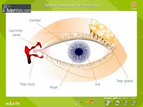
External features of The Human Eye - YouTube
Label the Eye Worksheet - Teacher-Made Learning Resources - Twinkl The first page is a labelling exercise with two diagrams of the human eye. One is a view from the outside, and the other is a more detailed cross-section. On the second page, you'll find a set of answers showing the properly labelled human eyes, designed to help you check the worksheets without having to come up with your own answer key.

Normal histology of Retina Eye Pathology Online - | Human anatomy and ...
Eye labeling Diagram | Quizlet a ring of muscle tissue that forms the colored portion of the eye around the pupil and controls the size of the pupil opening Cornea The clear tissue that covers the front of the eye Posterior Compartment filled with vitreous humor Pupil opening in the center of the iris Susponsory Ligament Allows the eye to move up and down Related questions
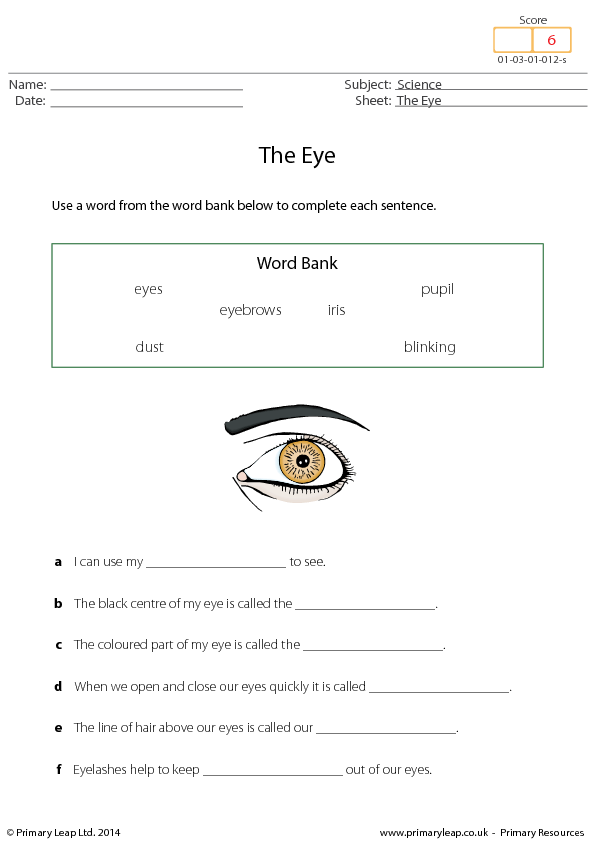
The Eye- Missing Words
Labeled Eye Diagram | Science Trends The Iris The iris is a structure found in the eyes of most mammals, and along with the pupil it controls how much light enters the eye. The iris is comprised of two different layers: the stroma, and the pigmented epithelial cells. The upper layer, the stroma, is linked to muscles that contract and dilate the pupil.

Draw a labeled diagram of human eye Write the functions of Cornea, Iris ...
The Eye - diagram to label | Teaching Resources File previews. pdf, 2.94 MB. Diagram of eye with key words to use in labelling it. Tes classic free licence.

Labeled Parts of the Rabbit | Pet rabbit care, Rabbit facts, Rabbit ...
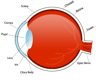
Eye Diagram With Labels and detailed description - BYJUS A brief description of the eye along with a well-labelled diagram is given below for reference. Well-Labelled Diagram of Eye The anterior chamber of the eye is the space between the cornea and the iris and is filled with a lubricating fluid, aqueous humour. The vascular layer of the eye, known as the choroid contains the connective tissue.
picture front of the eye without labels clipart - Clipground
Labelled Diagram of Human Eye, Explanation and Function - VEDANTU The human eye is a part of the sensory nervous system. Labeled Diagram of Human Eye The eyes of all mammals consist of a non-image-forming photosensitive ganglion within the retina which receives light, adjusts the dimensions of the pupil, regulates the availability of melatonin hormones, and also entertains the body clock.
Biology diagram worksheet
Diagram of eye with labels Labelling the eye. Use this interactive to label different parts of the human eye. Drag and drop the text labels onto the boxes next to the diagram. Selecting or hovering over a box will highlight each area in the diagram. The coloured part of the eye with the pupil at the centre. Diagram showing the parts of the eye with labels.
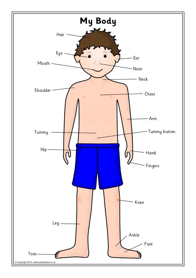
Body labelling
Human Eye Diagram, How The Eye Work -15 Amazing Facts of Eye The shark has even been used in human eye surgery! FACT 4 The length of our eyes are about 1 inch across and weigh about 0.25 ounce. FACT 5 Our eyeballs stay the same size forever but our nose and ears continue to grow. FACT 6 Eyes are the second most complex organ after the brain.
Parts of Human Eye and Their Functions | MD-Health.com
PDF Eye Anatomy Handout - National Eye Institute of light entering the eye. Lens: The lens is a clear part of the eye behind the iris that helps to focus light, or an image, on the retina. Macula: The macula is the small, sensitive area of the retina that gives central vision. It is located in the center of the retina. Optic nerve: The optic nerve is the largest sensory nerve of the eye.
Post a Comment for "45 eye diagram with labelling"