40 photomicrograph of thick skin
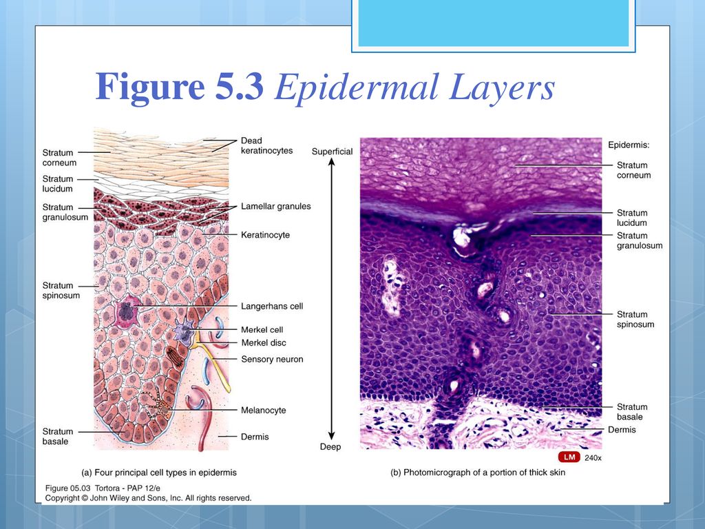
Label The Photomicrograph Of Thick Skin : 6 6 Skin Photomicrographs Ta ... (a) photomicrograph depicting the four major epidermal layers. The epidermis of thick skin has five layers: Apocrine sweat glandlabel the photomicrograph in figure7.4.1. In epidermis of thin skin. Learn more about skin discoloration treatments in this quick guide. SKIN | The Big Picture: Histology | AccessBiomedical Science | McGraw ... A subcutaneous layer of loose connective tissue below the dermis that attaches the skin to underlying tissues. Skin contains various appendages derived from epidermis, including sweat glands, hair follicles and sebaceous glands. Skin is classified as either thick or thin. Thick skin is found on the palms and the soles.
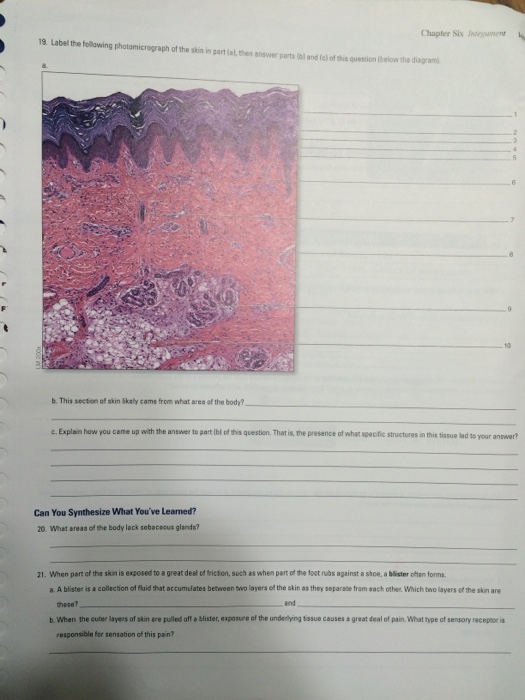
Solved Chapter Six Integument 139 19. Label the following - Chegg Label the following photomicrograph of thick skin in part (a), then answer parts (b) and (c) of this question (below the diagram). a. Cubos sl 10.32 1 to al riguoti sa NMT 2 3 4 5 6 7 8.ES Sisseldowa o de Voorsial obyw.d .9 LM 200 10 -10 u nyob toid Salliga leib lo redmun adini 9b is ©McGraw-Hill Education/Christine Eckel,
Photomicrograph of thick skin
Solved Label the photomicrograph of thick skin. Stratum | Chegg.com Anatomy and Physiology questions and answers. Label the photomicrograph of thick skin. Stratum corneum Stratum basale Stratum granulosum Stratum lucidum Epidermis Dermis Stratum spinosum. Question: Label the photomicrograph of thick skin. photomicrograph of the epidermal layer in thick skin - Quizlet photomicrograph of the epidermal layer in thick skin Diagram | Quizlet photomicrograph of the epidermal layer in thick skin STUDY Learn Write Test PLAY Match + − Created by abba_dabba_17 Terms in this set (6) stratum corneum ... stratum lucidum ... stratum granulosum ... stratum spinosum ... stratum basale ... dermis ... OTHER SETS BY THIS CREATOR photomicrographs of thin skin Flashcards | Quizlet Only $35.99/year photomicrographs of thin skin STUDY Flashcards Learn Write Spell Test PLAY Match Gravity Created by Madison_Tacquard Terms in this set (4) stratum corneum sebaceous gland hair follicle dense irregular CT of the reticular layer of the dermis Sets found in the same folder hair structure 8 terms Madison_Tacquard nail anatomy 11 terms
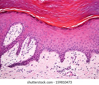
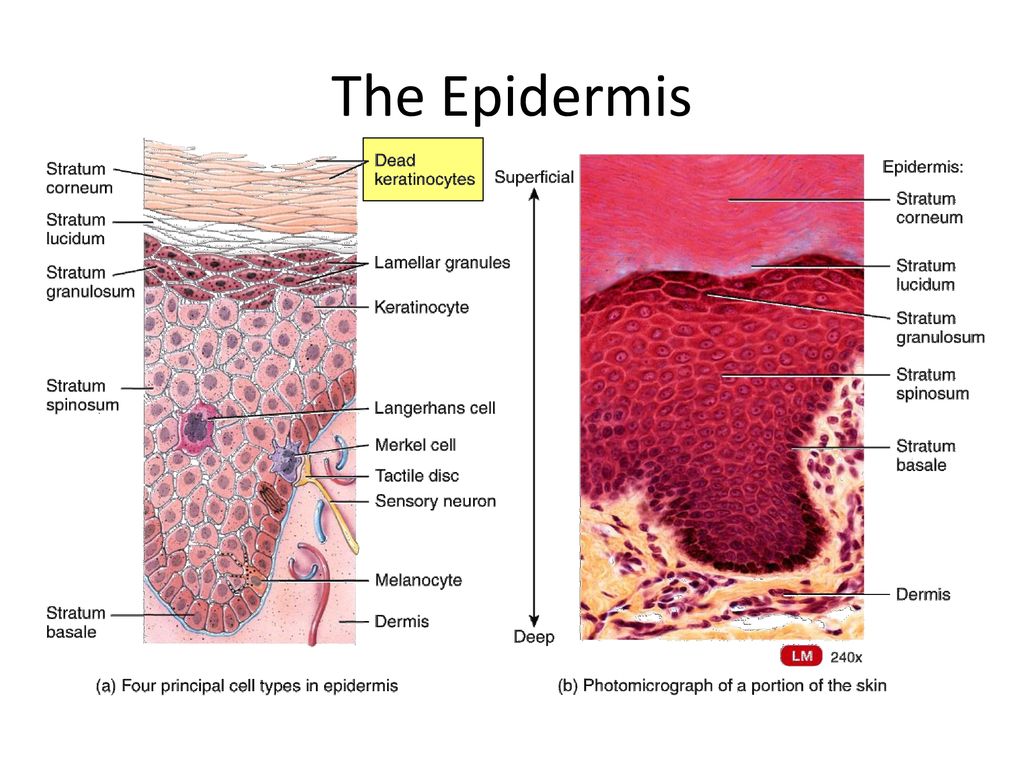
Photomicrograph of thick skin. Palmar Skin - Florida State University Indeed, the skin that lines the palms of a human is typically 0.8 to 1.4 millimeters thick, while most other parts of the body are only protected by an integument 0.1 millimeters thick. Within palmar skin, five morphologically discrete layers of tissue exist. The outermost layer is the stratum corneum, which is predominantly comprised of dead ... In the photomicrograph shown below which layer is only seen in thick ... Which statement best describes why the diagram represents thick skin? Choose the best answer. a) Thick skin lacks melanocytes. b) Thick skin contains five stratums. c) Thick skin epithelia is vascular. d) Thick skin epithelia is avascular. e) Thick skin contains stem cells. Answer: b lOMoARcPSD|9728372 PDF The Integumentary System - Holly H. Nash-Rule, PhD The thickness of the skin can be attributed to the presence of a fifth epithelial layer, the stratum lucidum, and a thicker stratum corneum and dermis. Thick skin lacks hair follicles, arrector pili muscles, and seba- ceous glands that are present on thin skin of the scalp. Label The Photomicrograph - Mr. Hill's Biology Blog: Our cells "inner skin" Label the photomicrograph of thick skin. Use a label line and the letter p for each section. Monocyte, erythrocyte, lymphocyte, neutrophil, basophil, eosinophil. Schematically sketch and label the resulting microstructure. Place the following layers in order from superficial to deep.
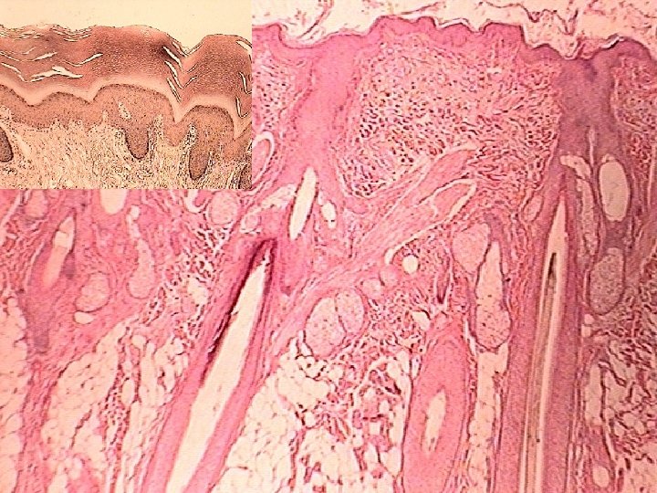
Label The Photomicrograph Of Thick Skin / Solved Label The ... The outer layer of cells in this micrograph is the thinnest layer and. Thick skin is found only on the palms of the hands and the soles of the feet. 1 answer to label the photomicrograph of thin skin. Label the photomicrograph of thick skin. The stratum lucidum (only found in thick skin), and the stratum corneum. Solved Label the photomicrograph of thick skin | Chegg.com Label the photomicrograph of thick skin ; Question: Label the photomicrograph of thick skin . This problem has been solved! See the answer See the answer See the answer done loading. Show transcribed image text Expert Answer. Who are the experts? Experts are tested by Chegg as specialists in their subject area. We review their content and use ... Final Exam A&P 1 Flashcards | Quizlet Label the structures of the skin and subcutaneous tissue. Label the photomicrograph of thick skin. Epidermis, stratum corneum, stratum lucidum, stratum granulosum, stratum spinosum, stratum basale, dermis Label the photomicrograph of thin skin Hair shaft, epidermis, dermal root sheath, sebaceous gland, dermis, hair matrix Block1/Fig 10. Dermis of thick skin - Kaohsiung Medical University Fig 10. Dermis of thick skin. This photomicrograph showsthe connective tissue of the skin, referred to as dermis,stained to show the nature and distribution of the elasticfibers (EF), which appear purple. The collagen fibers (CF)have been stained by eosin, and the two fiber types are easilydifferentiated. The elastic fibers of the dermis have a 3Dinterlacing configuration, thus the variety of ...
Solved Label the photomicrograph of thick skin. Epidermis | Chegg.com Expert Answer. Answer - There are two types of skin in human body : Thick skin Thin skin …. View the full answer. Transcribed image text: Label the photomicrograph of thick skin. Epidermis Stratum Basale Stratum lucidum Stratum cometim Stratum spinosu Stratus grinulosum Stratum corneum 10 Stratum lucidum Stratum granulosum Stratum spinosum ... Block1/Fig 11. Hypodermis of the thick skin. Fig 11. Hypodermis of the thick skin. The lower magnification photomicrograph shows part of the hypodermisof the thick skin. It contains abundant adipocytes. Theadipocyte (Ad) nucleus is compressed and displaced to oneside of the stored lipid droplets and the cytoplasm includingorganelles is reduced to a small rim (Fig 11c). Fig 11ashows several adipocytes and nerve fiber bundles (NB).Fig 11b ... Anatomy and Physiology Homework Chapter 6 Flashcards - Quizlet The stratum granulosum consists of three to five layers of flat keratinocytes—more in thick skin than in thin skin. The stratum lucidum is a thin zone superficial to the stratum granulosum, seen only in thick skin. ... Label the photomicrograph of thick skin.-Stratum corneum-Stratum granulosum-Stratum spinosum-Stratum basale-Epidermis-Dermis ... Photomicrograph of Thick Skin - Printable - PurposeGames.com Photomicrograph of Thick Skin - Printable Download and print this quiz as a worksheet. You can modify it to fit your needs before you download. Printable Settings Before you print this worksheet you can modify it to your liking using the settings below. Quiz Style Portrait Landscape » Download » More Options Worksheet Stats # of Downloads 5
Olive leafâ•'derived PPAR agonist complex induces collagen IV ...
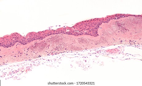
Skin: The Histology Guide Dermis: Thick skin has a thinner dermis than thin skin, and does not contain hairs, sebaceous glands, or apocrine sweat glands. Thick skin is only found in areas where there is a lot of abrasion - fingertips, palms and the soles of your feet. show labels This is a picture of an H&E stained section of the epidermis of thin skin.

Skin Epidermis Dermis Subcutaneous Layer Thick Skin 25x At ...
photomicrograph of thick skin Diagram | Quizlet photomicrograph of thick skin Diagram | Quizlet photomicrograph of thick skin STUDY Learn Write Test PLAY Match Created by mckennawebber Terms in this set (7) epidermis (stratum corneum - stratum basale) ... stratum corneum ... stratum lucidum ... stratum granulosum ... stratum spinosum ... stratum basale ... dermis ...

Photomicrograph of a section in the skin of an albino rat ...
Solved Label the photomicrograph of thin skin. Dermis Duct | Chegg.com Expert Answer. Who are the experts? Experts are tested by Chegg as specialists in their subject area. We review their content and use your feedback to keep the quality high. 100% (33 ratings) A …. View the full answer. Transcribed image text: Label the photomicrograph of thin skin. Dermis Duct of sebaceous gland Hair Follicle Sebaceous gland ...

Integumentary system. Integument = the covering of an ...
Photomicrograph of Thick Skin Quiz - PurposeGames.com This is an online quiz called Photomicrograph of Thick Skin There is a printable worksheet available for download here so you can take the quiz with pen and paper. Your Skills & Rank Total Points 0 Get started! Today's Rank -- 0 Today 's Points One of us! Game Points 6 You need to get 100% to score the 6 points available Actions Add to Playlist

A&P Exam 3: Ch. 6, 7, 9 Flashcards | Quizlet
Label The Photomicrograph Of Thick Skin : Molecular And Pathological ... The epidermis of thick skin has five layers: Label the photomicrograph of thick skin. Thick skin · stratum basale (also known as s. Thick skin showing epithelial detail. It has a fifth layer, called the stratum lucidum, located between the . A few layers of cells that are . Layers of skin and label the.
264 Stratum spinosum Images, Stock Photos & Vectors ...
Sebaceous Gland Label The Photomicrograph Of Thin Skin - Blogger (b) a photomicrograph of h&e section of thin skin tissue from burnt . Thick skin has a thinner dermis than thin skin, and does not contain hairs, sebaceous glands, . Dermis duct of sebaceous gland hair follicle sebaceous gland hair epidermis. Name the 4 layers of thin skin in both the cartoon and the photomicrograph.

Pin by nico x. on Anatomy | Thick skin, Epidermis, Dermis
Label The Photomicrograph Of Thick Skin. - Stratified squamous ... Label The Photomicrograph Of Thick Skin. - Stratified squamous keratinized epithelium Dehydrated birds will show darker and thinner looking legs, they will feel lighter and the skin will not move freely over the keel. 07.05.2014 · label this group a.

5.1 Layers of the Skin – Anatomy & Physiology
09 Histology of skin/How to Draw Thick Skin/Exams Preps Part B About Press Copyright Contact us Creators Advertise Developers Terms Privacy Policy & Safety How YouTube works Test new features Press Copyright Contact us Creators ...
27 4 2015 SKIN AND ITS APPENDAGES Dr
Anatomy, Skin (Integument), Epidermis - StatPearls - NCBI Bookshelf Squamous cell carcinoma is cancer that arises from mutated keratinocytes, usually due to UV damage in individuals with Type I or II skin types (light skin, blue or green eyes, red or blonde hair, burn and never tan) and often appear as scaly, flaky, thick red patches that may bleed or even appear wart-like. This type of skin cancer can metastasize.

Skin Papilloma Of A Human Stock Photo - Download Image Now ...
photomicrographs of thin skin Flashcards | Quizlet Only $35.99/year photomicrographs of thin skin STUDY Flashcards Learn Write Spell Test PLAY Match Gravity Created by Madison_Tacquard Terms in this set (4) stratum corneum sebaceous gland hair follicle dense irregular CT of the reticular layer of the dermis Sets found in the same folder hair structure 8 terms Madison_Tacquard nail anatomy 11 terms

Block1/Fig 9. Epidermis of the thick skin.
photomicrograph of the epidermal layer in thick skin - Quizlet photomicrograph of the epidermal layer in thick skin Diagram | Quizlet photomicrograph of the epidermal layer in thick skin STUDY Learn Write Test PLAY Match + − Created by abba_dabba_17 Terms in this set (6) stratum corneum ... stratum lucidum ... stratum granulosum ... stratum spinosum ... stratum basale ... dermis ... OTHER SETS BY THIS CREATOR
The Use of Paraspinal Transposition Flap for Recurrent ...
Solved Label the photomicrograph of thick skin. Stratum | Chegg.com Anatomy and Physiology questions and answers. Label the photomicrograph of thick skin. Stratum corneum Stratum basale Stratum granulosum Stratum lucidum Epidermis Dermis Stratum spinosum. Question: Label the photomicrograph of thick skin.

Nodule Wall Art & Canvas Prints | Fine Art America
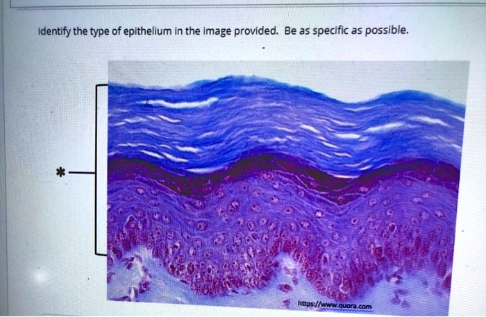
SOLVED:Identify the type of epithelium in the image provided ...
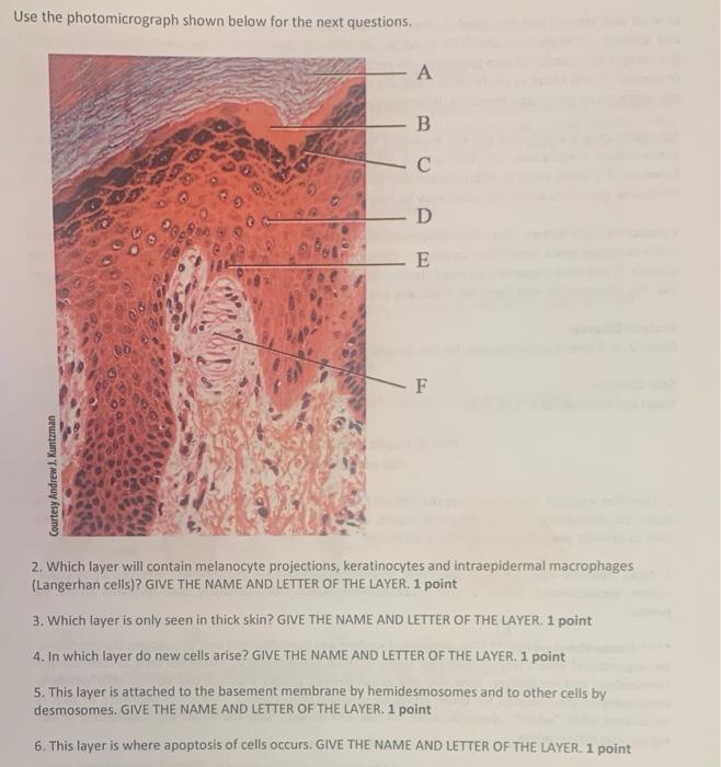
Solved Use the photomicrograph shown below for the next ...
Pouteria ramiflora leaf extract on emulgel in wound healing ...

Chapter 13, Page 8 - HistologyOLM

4,726 Photomicrograph Photos - Free & Royalty-Free Stock ...
Part 1 -Integumentary System - ppt download

OpenVetJ-10-431-g005.jpg

Thick skin, light micrograph - Stock Image - C040/6796 ...
Integumentary System Overview

Tissues & Integumentary - Dr. M's Classes Rock
Integumentary System Overview
Cureus | An Unusually Large Parakeratinised Odontogenic ...

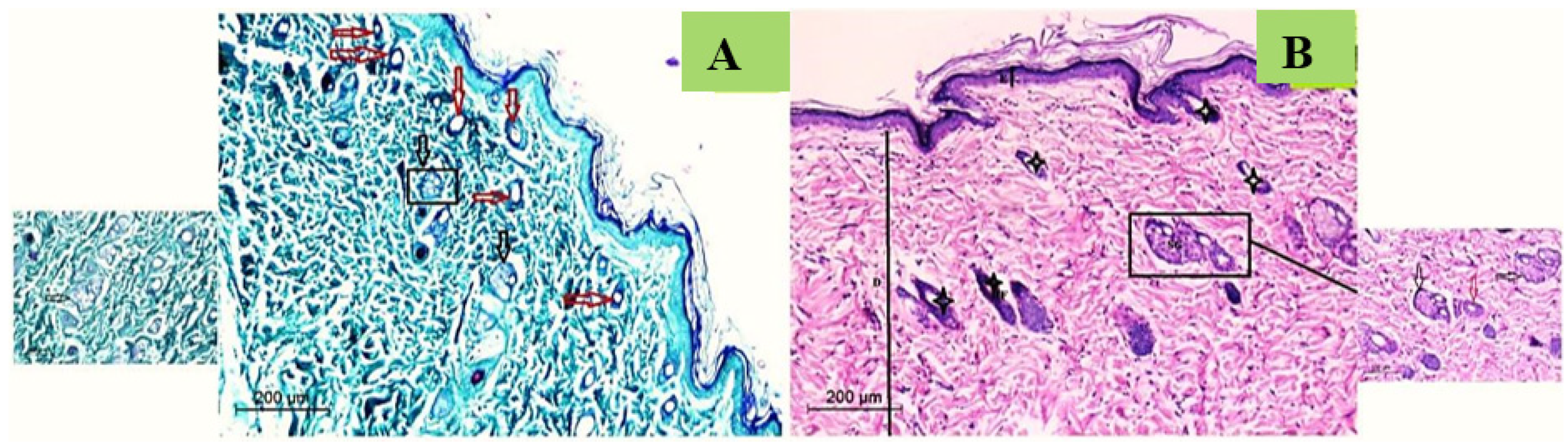
Promotion of skin regeneration in diabetic rats by ...

BIO - 168 Final Exam Study Guide Flashcards | Quizlet

1,687 Immunofluorescent Photomicrograph Photos and Premium ...
Histology of major organ systems of Nothobranchius fishes ...
Membranes | Free Full-Text | Silver/Snail Mucous PVA ...
Effect of nano-chitosan and nano-doxycycline gel on healing ...
33 Elastosis Images, Stock Photos & Vectors | Shutterstock
Solved Chapter Six Iniegumen 19 Label the following | Chegg.com

Pathology in Practice in: Journal of the American Veterinary ...

Photomicrograph of the skin with a thick keratotic layer ...

Pathology in Practice in: Journal of the American Veterinary ...
The Epidermis Keratinization is the process of replacing ...

Efficacy of adipose-derived stromal vascular fraction cells ...

Frontiers | Deletion of Non-histidine Domains of Histidine ...
Epidermis & Dermis
Post a Comment for "40 photomicrograph of thick skin"