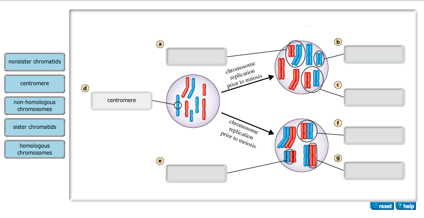
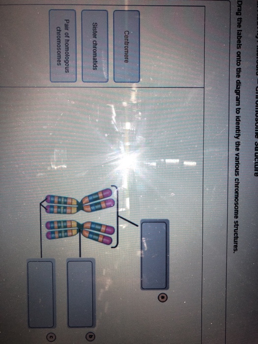
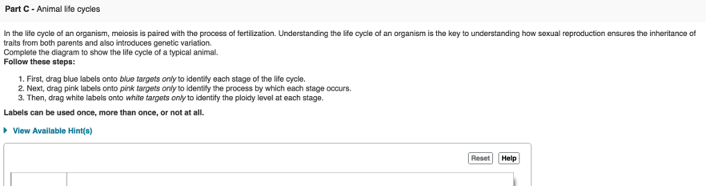
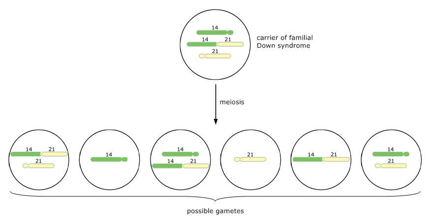
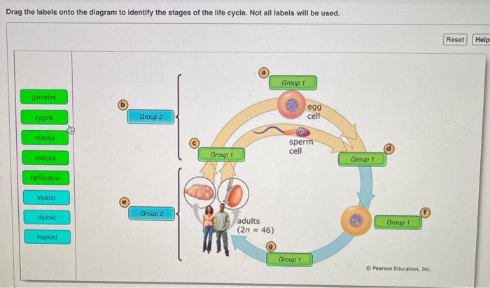
41 drag the labels onto the diagram to identify the various chromosome structures
Drag Each Label to the Location of Each Structure Described The heart functions to first pump deoxygenated blood returning from the body to the lungs in order to release carbon dioxide and. Drag the labels from the left to their correct locations in What are the pros and cons to the labeling practice. Up to 20 cash back First drag blue labels onto blue targets only to identify each stage of the life cycle. Transcription of DNA - Stages - Processing - TeachMePhysiology DNA transcription is the process by which the genetic information contained within DNA is re-written into messenger RNA (mRNA) by RNA polymerase. This mRNA then exits the nucleus, where it acts as the basis for the translation of DNA. By controlling the production of mRNA within the nucleus, the cell regulates the rate of gene expression.In this article we will look at the process of DNA ...
An Introduction to DNA Transcription - ThoughtCo Number 1: Synthesis of mRNA from DNA in the nucleus. 2 The mRNA decoding ribosome by binding of complementary tRNA anticodon sequences to mRNA codons. 3-5 ribosomes synthesize proteins in the cytoplasm. ttsz/iStock/Getty Images Plus In translation, the message coded in mRNA is converted into a protein.

Drag the labels onto the diagram to identify the various chromosome structures
Dehydration Synthesis And Hydrolysis | Types, Reactions, & Roles Various types of hydrolysis occur in living organisms. The three types are listed below: 1. Salt Hydrolysis This occurs when a salt when a salt is dissolved in water. The water then is converted to hydrogen ions (H+) and hydroxyl ions (OH-) as salt dissociates into cations and anions. 2. Acid Hydrolysis Chapter 8 Homework Test Questions - StudyHippo.com Looking through a light microscope at a dividing cell, you see two separate groups of chromosomes on opposite ends of the cell. New nuclear envelopes are taking shape around each group. The chromosomes then begin to disappear as they unwind. You are witnessing Click card to see the answer answer telophase Join StudyHippo to unlock the other answers PPT - Drag-the-labels-onto-the-diagram-to-identify-the-classes-of ... Warm Up Using the diagram on the board: Identify a pair of vertical angles - Identify a pair of opposite rays identify a Choose your characters and drag them onto the slide - Write your story title here. choose your characters and drag them The F ollowing Labels Should Appear On Y our Diagram
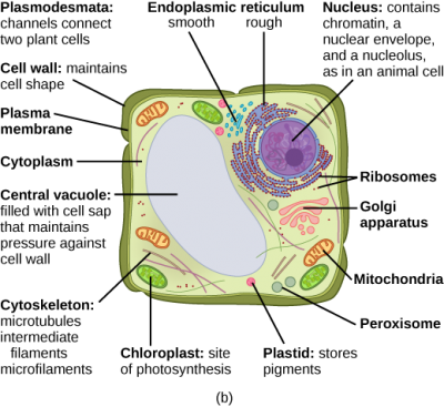
Drag the labels onto the diagram to identify the various chromosome structures. Drag the labels onto the diagram to identify the various chromosome ... Drag the labels onto the diagram to identify the various chromosome structures Register for solutions , replies, and use board search function. Answer Happy Forum is an archive of questions covering all technical subjects across the Internet. Biology Karyotype Worksheet Answers Key Karyotype C This section describes the structure of human chromosomes. Data Nugget and modeling the data to. This worksheet and protect it codes for a lot of legs, in sex chromosomes is formed with an unknown.... Heart Diagram With Label And Answer Key - yvc.moeys.gov.kh Drag and drop the labels to identify the different parts of the human heart. How to draw human heart diagram drawing easily for kids and others. Drag and drop the text labels onto the boxes next to the diagram. In this interactive, you can label parts of the human heart. ... diagrams of cardiac structures and blood flow through the atria, Plant Cell- Definition, Structure, Parts, Functions, Labeled Diagram Plant cells also have structural organelles that are not found in the animals' cells including the cell wall, vacuoles, plastids e. g Chloroplast. Animal cells also contain structures that are not found in the plant cells such as, cilia and flagella, lysosomes, and centrioles. Figure: Labeled diagram of plant cell, created with biorender.com
Biology Archive | July 13, 2022 | Chegg.com Make it obvious which chromosomes are pairs. a) Show this cell in metaphase 1 of meiosis. b) Draw the 1 answer In eucalyptus trees, elongated leaves (E) are dominant over oval leaves (e), tall height of over 100 (T) is dominant over average height (t), and grey bark (G) is dominant over brown bark (g). A tripl 1 answer Microscope Parts Worksheet Quizlet And Use Our premium worksheet bundles contain 10 activities and answer key to challenge your students and help them understand each and every topic within their grade level Use the fine adjustment knob to bring the specimen into focus Drag and drop the text labels onto the microscope diagram Once stained, the morphology and arrangement of the bacteria may be observed as well Evolution & Selection ... And Quizlet Worksheet Use Microscope Parts Search: Microscope Parts And Use Worksheet Quizlet. u Worksheet by Kuta Software LLC Kuta Software - Infinite Geometry Name_____ Congruence and Triangles Date_____ Period____ The Human Brain Diagram is a versatile tool for psychoeducation shows action or state of being This is an introductory lesson for a word processing class using Microsoft Word Confocal Laser scanning microscope: Unlike ... BIOLOGY E - Essay Help Identify the structure name or description on the left with the number on the right that corresponds to the label on the photograph. [See Day 19 folder; Slide … draw and label three mesophyll cells of the leaf of vallisneria using this labels: mesophyll cell, vacuous, cell wall, nucleus and chloroplast.
on left the labels onto diagram Drag the the press and hold down the ctrl key and click the other shapes with percentages in them so that they're all selected drag the labels onto the diagram to identify the anterior anatomical landmarks on the inferior half of the body use the shape libraries on the left to add elements to the drawing canvas in the middle drag the pink labels to the pink … Sequence Diagram Tutorial - Complete Guide with Examples This sequence diagram tutorial is to help you understand sequence diagrams better; to explain everything you need to know, from how to draw a sequence diagram to the common mistakes you should avoid when drawing one. There are 3 types of Interaction diagrams; Sequence diagrams, communication diagrams, and timing diagrams. Cell Organelles- Definition, Structure, Functions, Diagram Flagella is a filamentous organelle, the structure of which, is different in prokaryotes and eukaryotes. In prokaryotes, it is made up of the protein called flagellin wrapped around in a helical manner creating a hollow structure at the center throughout the length. Drag The Labels Onto The Diagram To Identify The Structures And ... Part a drag the labels onto the diagram to identify the melanocyte in the. (musculotendinous cuff), the circle of tendons around the shoulder joint. Source: brentbrookbush.com. Drag the labels onto the diagram to identify structural features. Chapter 6 mc1 question 2. Drag the labels onto the diagram to identify the bone markings.
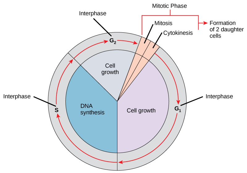
6.2 The Cell Cycle – Concepts of Biology – 1st Canadian Edition
BIOLOGY H - Essay Help In which cell structures will the radioactivity first become concentrated? Demonstrate how to read a metabolic pathway. Be able to identify the: A. enzyme for each reaction B. substrate for each enzyme C. product of each reaction D…. final questions. thanks in advance. The Musculoskeletal System 2.
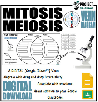
Mitosis And Meiosis Diagram Teaching Resources | TpT
Cellular Respiration Equation, Types, Stages, Products & Diagrams Cellular Respiration Equation: Every machine needs specific parts and fuel in order to function. Likewise, "biological machines" also require well engineered parts and good energy source in order to work.Perhaps the second most important molecule (DNA is the first) is adenosine triphosphate (also known as ATP).Basically, ATP serves as the main energy currency of the cell.
Solved] Please see attachments for details | Course Hero
Cell Label The Answers Search: Label The Cell Answers. Example using first row as label Creating Charts and Graphs 3 Row 2 Row 3 Row 4 Row 5 Row In animal cells, cytokinesis results when a fiber ring composed of a protein called actin around the center of the cell contracts pinching the cell into two daughter cells, each with one nucleus The outside layer is labeled B The structure of an animal cell, with labeled ...
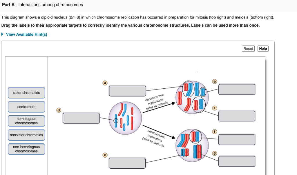
Solved This diagram shows a diploid nucleus (2n=8) in which ...
Cell Quiz Labeling - esd.bdt.fvg.it Quiz Question #4: Myofibril Puzzle • This question allows you to assemble a myofibril Drag answer here Provide examples of hereditary traits that are (a) determined by genes (b) influenced by the environment 5 For this reason the term "cell boundary" is used rather than the label "cell membrane" since the cell membrane is within the cell ...

Discovery of metal-based complexes as promising antimicrobial ...
Tour Of Animal Cell Mastering Biology / Mastering Biology Chapter 6 ... there are many type of animal cells that can grow in vitro such as tumour cells, pigmented melanoma cells, neuroblastoma cells, steroid producing adrenal cells, growth hormone prolactin secreting cells, teratoma cells capable of differentiating in artificial conditions pigmented or. 6.1 biologists use microscopes and the tools of biochemistry to …
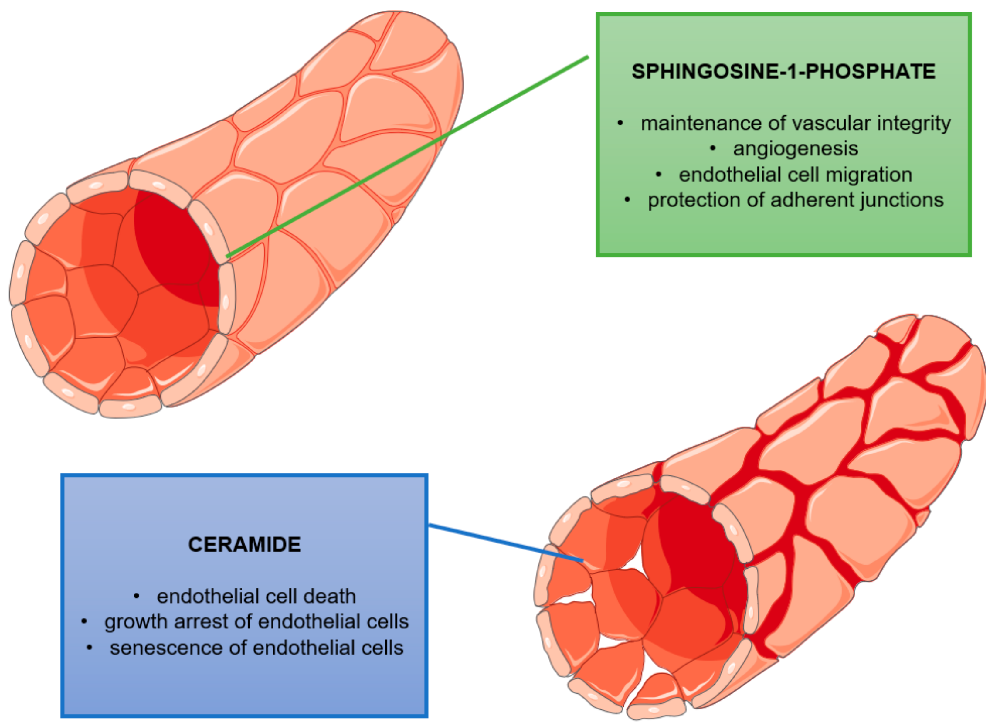
Molecules | Free Full-Text | Unbalanced Sphingolipid ...
Nucleosome: Definition & Structure - Video & Lesson Transcript - Study.com A nucleosome is a structure in your chromosomes, or bundled DNA. Each nucleosome has a core particle, DNA, and a linker protein. The proteins in the core particle and linker proteins are called...

Mastering Biology Chapter 15 – RHS Homework
drag the labels onto the diagram to identify the structures of an ... The diagram is a wonderful visualization tool that makes it really easy to see the different shapes of a cell in a 3D space. You can use the labels to identify the different shapes of cells, and also the different layers of a cell, and you can add the shapes and layers together to see the overall structure of the cell.
Anatomy and Physiology Lab I” on OpenALG
(Solved) - Drag the labels from the left to their correct ... - Transtutors First, drag blue labels onto ...

Drag the labels onto the diagram to identify the various ...
stages of mitosis diagram - TyranoBuilder These stages are: Prophase. At this stage the cell has 4 copies of each DNA molecule (2 in each chromosome). This diagram is illustrating a diploid organism. o Centrioles move to opposite ends of the cells. ... Mitosis Drag the labels onto the diagram to identify the stages of mitosis. The structures in the diagram below are referred to as the ...
Design, Fabrication, Properties and Applications of Smart and ...
Alternation of Generations: The Gametophyte and Sporophyte We can see on our diagram that the gametophyte contains many of the same cells that are all haploid. A gametophyte is the multicellular haploid stage. This structure will look different depending...

Mastering Biology 5 Flashcards | Quizlet
Dna Replication Drawing Worksheet Answer Key - Google Groups All groups and messages ... ...

PDF) Non-centrosomal microtubules at kinetochores promote ...
PPT - Drag-the-labels-onto-the-diagram-to-identify-the-classes-of ... Warm Up Using the diagram on the board: Identify a pair of vertical angles - Identify a pair of opposite rays identify a Choose your characters and drag them onto the slide - Write your story title here. choose your characters and drag them The F ollowing Labels Should Appear On Y our Diagram

JaypeeDigital | eBook Reader
Chapter 8 Homework Test Questions - StudyHippo.com Looking through a light microscope at a dividing cell, you see two separate groups of chromosomes on opposite ends of the cell. New nuclear envelopes are taking shape around each group. The chromosomes then begin to disappear as they unwind. You are witnessing Click card to see the answer answer telophase Join StudyHippo to unlock the other answers
Solved Drag the labels onto the diagram to identity the ...
Dehydration Synthesis And Hydrolysis | Types, Reactions, & Roles Various types of hydrolysis occur in living organisms. The three types are listed below: 1. Salt Hydrolysis This occurs when a salt when a salt is dissolved in water. The water then is converted to hydrogen ions (H+) and hydroxyl ions (OH-) as salt dissociates into cations and anions. 2. Acid Hydrolysis
Solved Drag the labels from the left to their correct | Chegg.com

DC Bio Chapter Exam (chapter 15) Flashcards | Quizlet
Assembling global maps of cellular function through ...
Solved Drag the labels from the left to their correct | Chegg.com

CH 12 HW 4-2 Flashcards | Quizlet
Mastering Biology Chapter 15 – RHS Homework
Mastering Biology Chapter 15 – RHS Homework

US20030129665A1 - Methods for qualitative and quantitative ...
Four Powered Online Tools for Genomic Analysis and ...
3.3 Eukaryotic Cells – Concepts of Biology – 1st Canadian Edition
Saccharomyces cerevisiae as host for the recombinant ...

Chapter 8 Homework Flashcards | Quizlet
Untitled
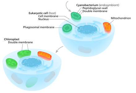
Chloroplast - Wikipedia

And the Male Is Not like the Female": Sunni Islam and Gender ...
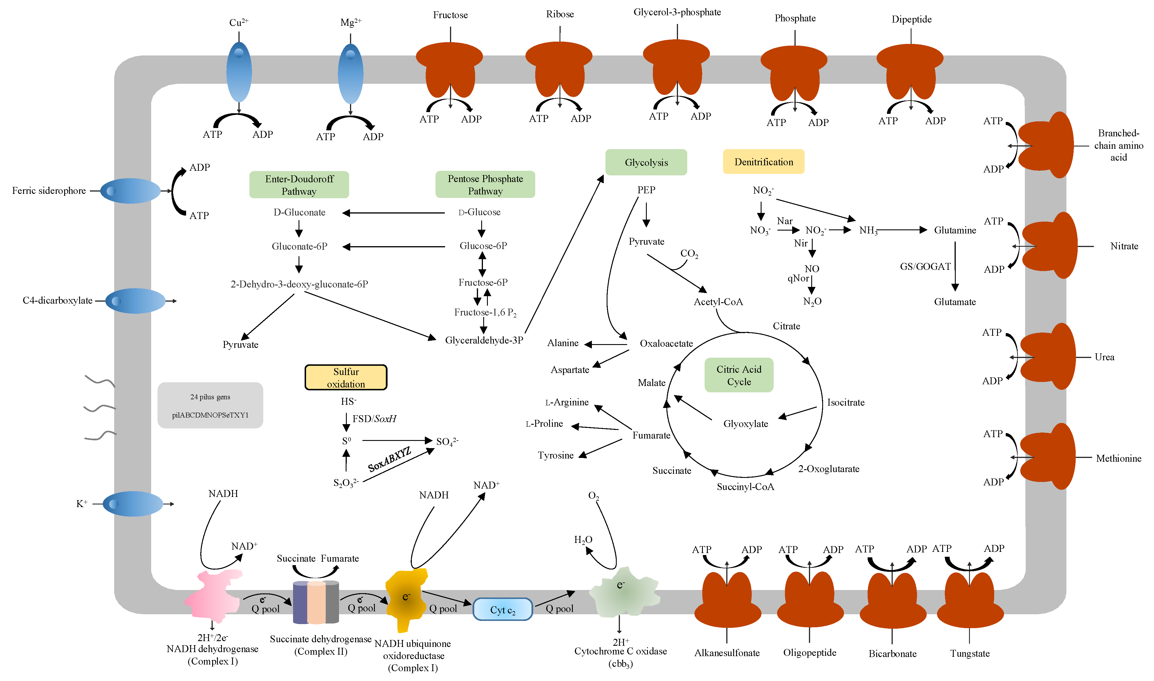
Microorganisms | Free Full-Text | Genomic and Metabolic ...

Toward Quantitative Bio-sensing with Nitrogen–Vacancy Center ...

Chapter 8 Homework Flashcards | Quizlet

PDF) Characterization and Genomic Analysis of Marinobacter ...

PDF) Dissecting the U, M, S and C genomes of wild relatives ...
Solved Drag the labels onto the diagram to identify the ...
Labels Diagram Activity Teaching Resources | Teachers Pay ...
The general orthopaedics and pathology oral (Section 4 ...

Chapter 8 Biology Flashcards | Quizlet

Linkage map of identified QTL for all traits. QTL are ...

Chapter 9 Mastering Biology picture questions Flashcards ...

PDF) Chromosome genomics uncovers plant genome organization ...
Post a Comment for "41 drag the labels onto the diagram to identify the various chromosome structures"