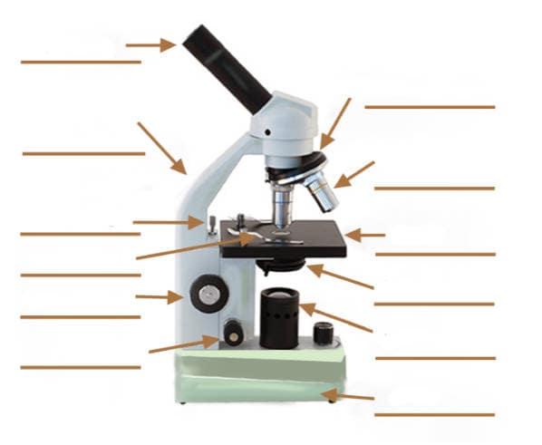
40 compound light microscope drawing with label
Home | Cedar Park Church Sunday Services. Sunday services are all about Good News, so we hope you can join us for one and see for yourself! All of our worship services include powerful music, practical teaching and preaching from the Bible, as well as opportunities for prayer and response, and time to build relationships with other people. Labeled microscope diagram - Pinterest A diagram showing all of the parts of a compound light microscope. Racing is really fun, especially if you can express your creativity through creating your own car. Here are the awesome pinewood derby templates that will inspire you and help you build a cool car easily. Art, Humor, & More!
Fiber Optic Terms and Definitions - Lightel Microscope Fiber Optic Inspection A microscope used to inspect the end surface of a connector for flaws or contamination or a fiber for cleave quality. Microsecond One millionth of a second or 10-6 seconds. Abbreviated µs. Microwatt One millionth of a Watt or 10-6 Watts. Abbreviated µW. MIL-SPEC Abbreviation for military specification.
Compound light microscope drawing with label
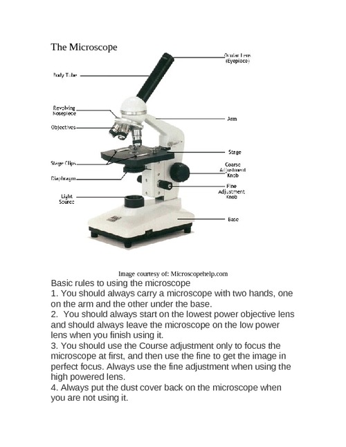
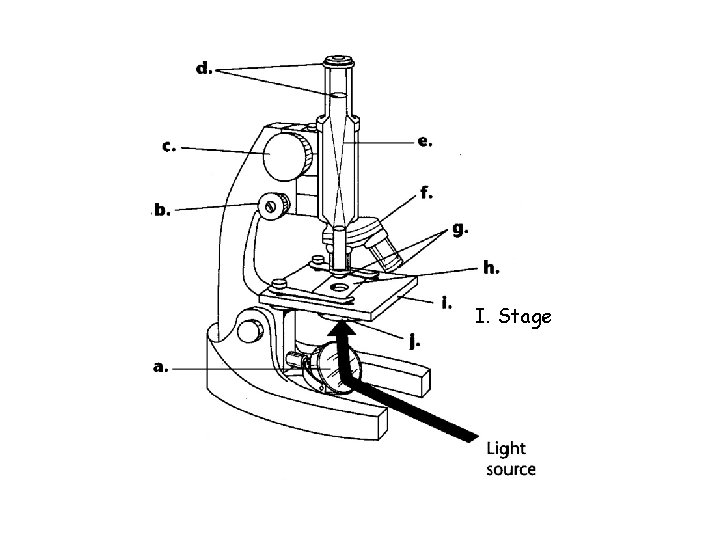
PDF The Compound Light Microscope - teachers.wrdsb.ca The Compound Light Microscope TASK Refer to page 605 in your text to: 1. Name each of the structures described in the table to the right. 2. Match each structure to the letter in the diagram below. ** ALWAYS USE TWO HANDS TO CARRY A MICROSCOPE** Letter Structure Function joins body tube to base supports the entire microscope COMPOUND LIGHT MICROSCOPE PARTS AND FUNCTIONS .pdf COMPOUND LIGHT MICROSCOPE Draw and label the parts of a compound light microscope. Parts and Functions of a compound light microscope. MAGNIFYING PARTS •EYEPIECE / OCULARS -the lens the viewer looks through to see the magnified specimen. Compound Microscope Parts - Labeled Diagram and their Functions Basically, compound microscopes generate magnified images through an aligned pair of the objective lens and the ocular lens. In contrast, "simple microscopes" have only one convex lens and function more like glass magnifiers. [In this figure] Two "antique" microscopes played significant roles in the history of biology.
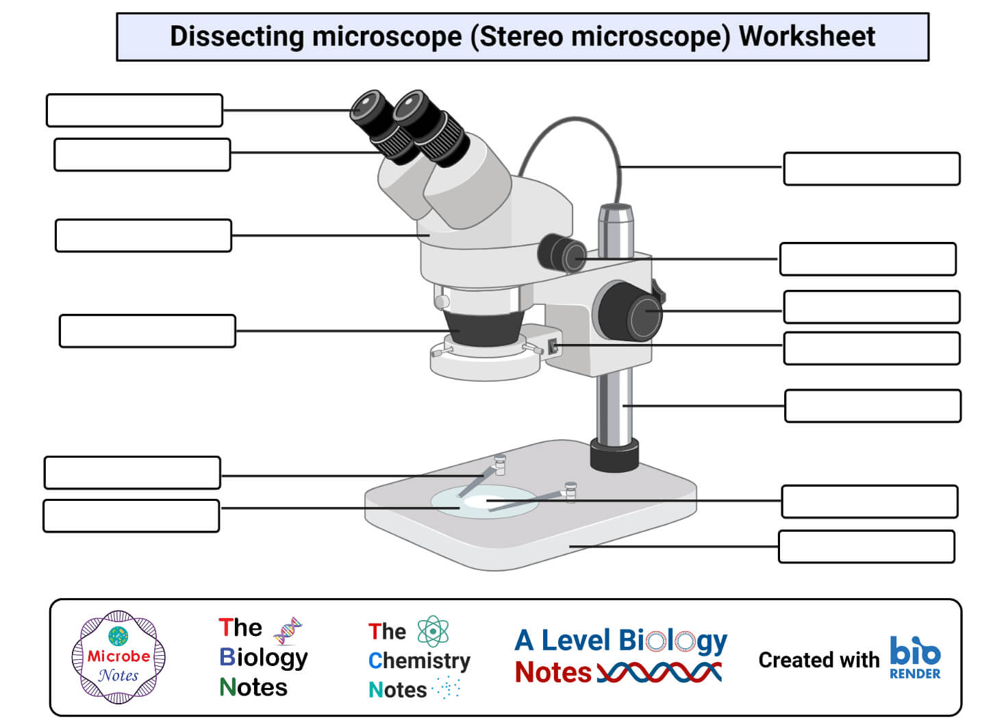
Compound light microscope drawing with label. Microscope Parts and Functions First, the purpose of a microscope is to magnify a small object or to magnify the fine details of a larger object in order to examine minute specimens that cannot be seen by the naked eye. Here are the important compound microscope parts... Eyepiece: The lens the viewer looks through to see the specimen. Compound Microscope: Definition, Diagram, Parts, Uses, Working ... - BYJUS A compound microscope is defined as A microscope with a high resolution and uses two sets of lenses providing a 2-dimensional image of the sample. The term compound refers to the usage of more than one lens in the microscope. Also, the compound microscope is one of the types of optical microscopes. The Compound Microscope.docx - The Compound Microscope 1. Draw and ... 2. Enumerate other different types of microscope and give their functions. TYPES OF MICROSCOPE FUNCTIONS 1. Stereo Microscope-provides a 3D image/ stereo image of the specimen.-magnification: 10x to 40x.-provides both transmitted and reflected illumination and can be used to view a sample that will not allow light to pass through it. 2. Compound Microscope-also known as Biological Microscope ... Cambridge Checkpoint Science Coursebook 8 - Academia.edu This captivating Coursebook provides coverage of stage 8 of the revised Cambridge Secondary 1 curriculum framework. It is endorsed by Cambridge International Examinations for use with their programme. The series is written by a highly experienced
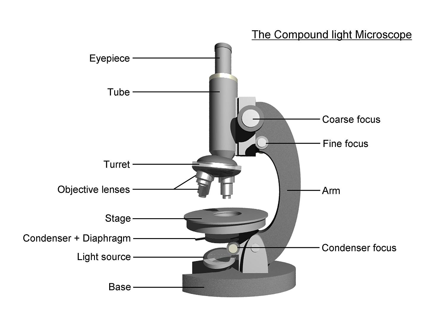
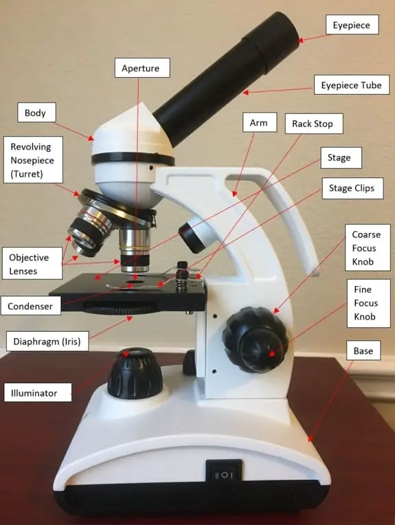
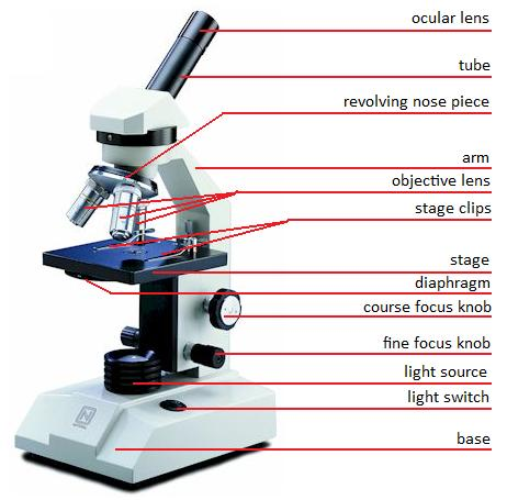
PDF Lab: Using a Compound Light Microscope When you use a compound light microscope, the specimen being studied is placed on a glass slide. The slide may be either a prepared slide that is permanent and was purchased from a science supply company, or it may be a wet mount that is made for temporary use and is made in the lab room. Objectives: In this lab you will: 1. Learn the parts of ... Compound Microscope - Diagram (Parts labelled), Principle and Uses Also called as binocular microscope or compound light microscope, it is a remarkable magnification tool that employs a combination of lenses to magnify the image of a sample that is not visible to the naked eye. Compound microscopes find most use in cases where the magnification required is of the higher order (40 - 1000x). Comparing the visible and the infrared types of light, which would … Ch. 4 - Prob. 10GRP Ch. 4 - Will the light given off by Earth’s surface easily... Ch. 4 - How does the total amount of energy coming from... Ch. 4 - What type of light primarily heats Earth’s surface... Ch. 4 - Is more energy absorbed by Earth’s surface in the... Ch. 4 - Due to the light absorbed by Earth’s surface that... Parts of a Compound Microscope and Their Functions - NotesHippo Compound microscope mechanical parts (Microscope Diagram: 2) include base or foot, pillar, arm, inclination joint, stage, clips, diaphragm, body tube, nose piece, coarse adjustment knob and fine adjustment knob. Base: It's the horseshoe-shaped base structure of microscope. All of the other components of the compound microscope are supported ...
UD Virtual Compound Microscope - University of Delaware ©University of Delaware. This work is licensed under a Creative Commons Attribution-NonCommercial-NoDerivs 2.5 License.Creative Commons Attribution-NonCommercial-NoDerivs 2.5 … Thioglycollate broth: Composition, Principle, and Uses 6.10.2016 · Thioglycollate broth is the enrichment broth most frequently used in diagnostic bacteriology. This broth supports the growth of anaerobes, aerobes, microaerophilic, and fastidious microorganisms. It contains many nutritive factors, including casein, yeast, beef extracts, and vitamins to enhance the growth of the most medically important bacteria. A Study of the Microscope and its Functions With a Labeled Diagram ... To better understand the structure and function of a microscope, we need to take a look at the labeled microscope diagrams of the compound and electron microscope. These diagrams clearly explain the functioning of the microscopes along with their respective parts. Man's curiosity has led to great inventions. The microscope is one of them. Solved Date Section Name EXERCISE 1 Laboratory Report: | Chegg.com Draw four fields of the specimens you observed. Label the objective power used and calculate the total magnification. If possible, show the same field or material at two different magnifications OOOO Specimen Objective lens Total magnification 2. On your This problem has been solved! See the answer Show transcribed image text Expert Answer
Parts of a Microscope and Their Functions
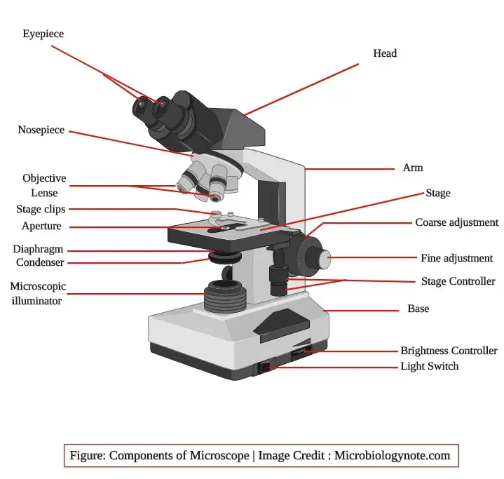
Parts of a microscope with functions and labeled diagram - Microbe Notes Microscopic illuminator - This is the microscopes light source, located at the base. It is used instead of a mirror. It captures light from an external source of a low voltage of about 100v. Condenser - These are lenses that are used to collect and focus light from the illuminator into the specimen.

Biology: The Compound Light Microscope Labeling Diagram | Quizlet
Compound Microscope- Definition, Labeled Diagram, Principle, Parts, Uses In order to ascertain the total magnification when viewing an image with a compound light microscope, take the power of the objective lens which is at 4x, 10x or 40x and multiply it by the power of the eyepiece which is typically 10x. Therefore, a 10x eyepiece used with a 40X objective lens will produce a magnification of 400X.

Parts of a Compound Microscope and Their Functions
Microscopy - Wikipedia Optical or light microscopy involves passing visible light transmitted through or reflected from the sample through a single lens or multiple lenses to allow a magnified view of the sample. The resulting image can be detected directly by the eye, imaged on a photographic plate, or captured digitally.The single lens with its attachments, or the system of lenses and imaging equipment, …

microscope drawing with label - Clip Art Library
GeM | Bidding Advanced search for Ongoing Bids. Search by Bid / RA Details; Search by Ministry / Organization; Bid / RA Number

Compound Microscope Parts – Labeled Diagram and their ...
Diagram of a Compound Microscope - Biology Discussion A bright-field or compound microscope is primarily used to enlarge or magnify the image of the object that is being viewed, which can not otherwise be seen by the naked eye. Magnification may be defined as the degree of enlargement of the image of an object provided by the microscope.

Can someone can send me diagram of this compound microscope ...
Compound Light Microscope Optics, Magnification and Uses In order to ascertain the total magnification when viewing an image with a compound light microscope, take the power of the objective lens which is at 4x, 10x or 40x and multiply it by the power of the eyepiece which is typically 10x. Therefore, a 10x eyepiece used with a 40X objective lens, will produce a magnification of 400X.

Parts of Microscope, Function, Names & Labeled Diagram ...
Solved EXERCISE 1 Laboratory Report: Introduction to the - Chegg EXERCISE 1 Laboratory Report: Introduction to the Compound Light Microscope Results 1. Draw four fields of the specimens you observed. Label the objective power used and calculate the total magnification. If possible, show the same field or material at two different magnifications. ОООО Specimen Objective lens Total magnification 2.

Modern Electronic Powerful Lab Microscope Parts Infographic ...
Compound Microscope Parts, Functions, and Labeled Diagram So, a compound microscope with a 10x eyepiece magnification looking through the 40x objective lens has a total magnification of 400x (10 x 40). Specimen or slide: The object used to hold the specimen in place along with slide covers for viewing. Most slides & slide covers are thin glass rectangles.
Simple Microscope - Diagram (Parts labelled), Principle ...
Labeling the Parts of the Microscope | Microscope activity, Science ... Jan 13, 2016 - Free worksheets for labeling parts of the microscope including a worksheet that is blank and one with answers. ... Description Worksheet identifying the parts of the compound light microscope. Answer key: 1. Body tube 2. Revolving nosepiece 3. Low power objective 4. Medium power objective 5. High power objective 6. Stage clips 7 ...
Compound Microscope Stock Illustrations – 727 Compound ...
How to draw compound of Microscope easily - step by step I will show you " How to draw compound of microscope easily - step by step "Please watch carefully and try this okay.Thanks for watching.....#microscopedrawi...

Compound Microscope Drawing With Parts and Functions
Parts of the Microscope with Labeling (also Free Printouts) Let us take a look at the different parts of microscopes and their respective functions. 1. Eyepiece it is the topmost part of the microscope. Through the eyepiece, you can visualize the object being studied. Its magnification capacity ranges between 10 and 15 times. 2. Body tube/Head It is the structure that connects the eyepiece to the lenses.

Label Microscope Diagram | Microscope parts, Microscope ...
anatomy of microscope Microscope labelled slides mitosis drawing parts slide drawings draw specimen properly using compound microscopy important lrm22 nau lessons edu notes Human Anatomy. 16 Pictures about Human Anatomy : Activity 1: Identifying the Parts of a Microscope | Anatomy and, Compound Microscope Reviews and Guide 2020 - TopMicroscopeReviews and also ...
Free Microscope Drawing, Download Free Microscope Drawing png ...
Compound Light Microscope: Everything You Need to Know A compound light microscope is a type of light microscope that uses a compound lens system, meaning, it operates through two sets of lenses to magnify the image of a specimen. It's an upright microscope that produces a two-dimensional image and has a higher magnification than a stereoscopic microscope.
Microscopy- History, Classification, Terms, Diagram
DePaul University | DePaul University, Chicago Our Commitment to Anti-Discrimination. DePaul University does not discriminate on the basis of race, color, ethnicity, religion, sex, gender, gender identity, sexual orientation, national origin, age, marital status, pregnancy, parental status, family relationship status, physical or mental disability, military status, genetic information or other status protected by local, state or federal ...
parts of a microscope - Clip Art Library
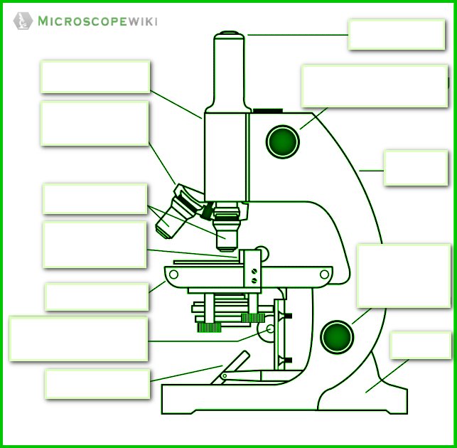
Label the microscope — Science Learning Hub Use this with the Microscope parts activity to help students identify and label the main parts of a microscope and then describe their functions. Drag and drop the text labels onto the microscope diagram. If you want to redo an answer, click on the box and the answer will go back to the top so you can move it to another box.

label microscope diagram | Charts | Microscope, Anatomy bones ...
Compound Light Microscope Labeling - Printable - PurposeGames.com This is a printable worksheet made from a PurposeGames Quiz. To play the game online, visit Compound Light Microscope Labeling Download Printable Worksheet Please note! You can modify the printable worksheet to your liking before downloading. Download Worksheet Include correct answers on separate page About this Worksheet
Compound Microscope Drawing - ClipArt Best
Light Microscope: Functions, Parts and How to Use It Compound light microscope: has a higher magnification than a simple microscope because it uses at least two sets of lenses, an objective lens and an eyepiece. Light Microscope Function. The function of the light microscope is based on its ability to focus a beam of light through a very small and transparent specimen, to produce an image.
simple light microscope labeled - Clip Art Library
Compound Microscope: Parts of Compound Microscope - BYJUS The parts of the compound microscope can be categorized into: Mechanical parts; Optical parts (A) Mechanical Parts of a Compound Microscope. 1. Foot or base. It is a U-shaped structure and supports the entire weight of the compound microscope. 2. Pillar. It is a vertical projection. This stands by resting on the base and supports the stage. 3. Arm

Optical microscope Drawing Worksheet, public space, angle ...
compound light microscopes - Teachers Pay Teachers 5. $2.25. PDF. The Compound Light Microscope is a great introductory lesson on the basics of the microscope and how it is used. This product includes a labeled color poster of the microscope and how to use it as well as worksheets that include reading comprehension, labeling, fill in the blank and a sequence card set.

Solved A. OLYMPUS C. B. Use the Diagram to answer the | Chegg.com
4.1. Chirality | Organic Chemistry 1: An open textbook - Lumen … We turn now to concept of chirality, discovered and explored by Louis Pasteur . The term chiral, from the Greek work for ‘hand’, refers to anything which cannot be superimposed on its own mirror image. Your hands, of course, are chiral – you cannot superimpose your left hand on your right, and you cannot fit your left hand into a right-handed glove (which is also a chiral object).

Compound Light Microscope Labels 1 (Champagne) Diagram | Quizlet
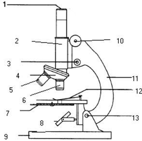
Labelled Diagram of Compound Microscope The concave surface is used without condenser and plain surface with condenser. (I) Magnification: To determine the magnification following formula is used: m = l/f × e ADVERTISEMENTS: Here m = magnification ADVERTISEMENTS: l = length of body tube = 160 mm e = magnification of eye piece. ADVERTISEMENTS: f = magnification of objective.

Parts of a Microscope - SmartSchool Systems
Compound Microscope Parts - Labeled Diagram and their Functions Basically, compound microscopes generate magnified images through an aligned pair of the objective lens and the ocular lens. In contrast, "simple microscopes" have only one convex lens and function more like glass magnifiers. [In this figure] Two "antique" microscopes played significant roles in the history of biology.

Photos of two standard compound microscopes: An inverted ...
COMPOUND LIGHT MICROSCOPE PARTS AND FUNCTIONS .pdf COMPOUND LIGHT MICROSCOPE Draw and label the parts of a compound light microscope. Parts and Functions of a compound light microscope. MAGNIFYING PARTS •EYEPIECE / OCULARS -the lens the viewer looks through to see the magnified specimen.

Cytology. Cytology. radiation used to illuminate the specimen ...
PDF The Compound Light Microscope - teachers.wrdsb.ca The Compound Light Microscope TASK Refer to page 605 in your text to: 1. Name each of the structures described in the table to the right. 2. Match each structure to the letter in the diagram below. ** ALWAYS USE TWO HANDS TO CARRY A MICROSCOPE** Letter Structure Function joins body tube to base supports the entire microscope
16 Parts of a Compound Microscope: Diagrams and Video ...
Cell Structure - Biology | Quiz

Parts of a microscope with functions and labeled diagram

Simple Microscope - Diagram (Parts labelled), Principle ...

Compound Microscope: Know Definition,working, diagram, properties
Parts of a microscope with functions and labeled diagram
microscope vector sketch 7312859 Vector Art at Vecteezy
Parts of the Microscope with Labeling (also Free Printouts ...

Labeling the Parts of the Microscope | Microscope activity ...

Compound Microscope Parts – Labeled Diagram and their ...

22 Parts Of a Microscope With Their Function And Labeled ...
Grade 10 Applied Science – Biology
Parts of a Compound Light Microscope Mirror Light
Drawing microscope - Teaching resources
Compound Light Microscope Labeled - ClipArt Best

Microscope Drawing Worksheet | Clipart library - Free Clipart ...
Post a Comment for "40 compound light microscope drawing with label"