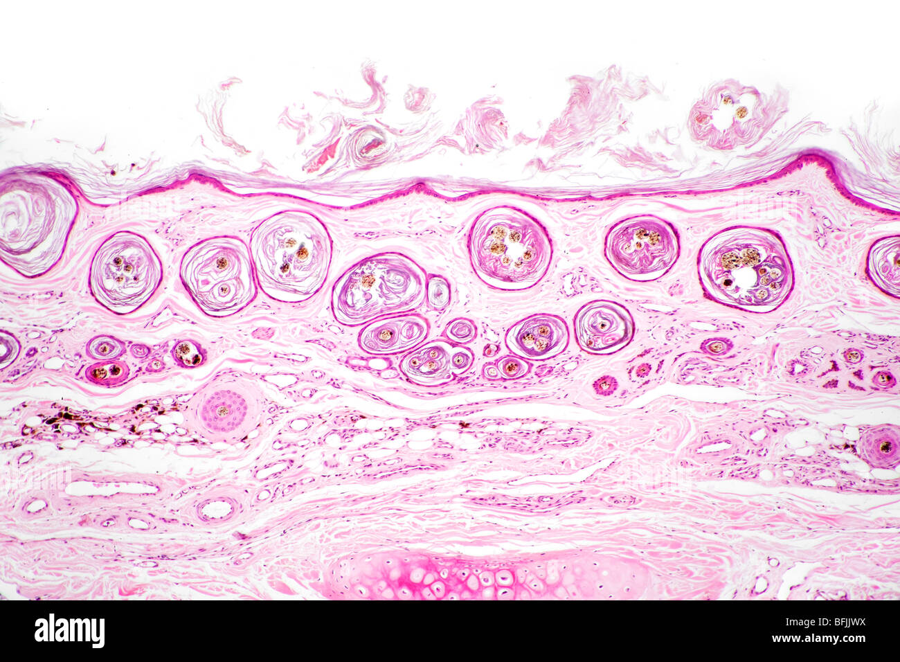
42 label the photomicrograph of the skin and its accessory structures.
(Solved) - Label The Photomicrograph Of The Skin And Its Accessory ... Possible answers:Pore, ... LibGuides: BIO 140 - Human Biology I - Textbook: Chapter 10 ... The epithelia lining the skin, parts of the mouth and nose, and the anus develop from the ectoderm. Cells lining the airways and most of the digestive system originate in the endoderm. The epithelium that lines vessels in the lymphatic and cardiovascular system derives from the mesoderm and is called an endothelium.
The Integumentary System Lab Report - 703 Words | Studymode A tan is your skin's attempt to prevent UV rays from doing any further damage to the sensitive skin cells in your epidermis. …show more content… Be sure to label all of the structures in the epidermis and dermis you were able to find: QUESTIONS: A. Compare your slide to the photomicrograph example in the lab procedure.
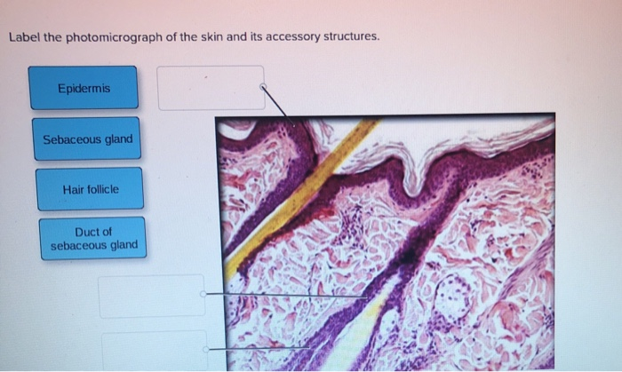
Label the photomicrograph of the skin and its accessory structures.
Label the photomicrograph in Figure 7.4. Examine a slide of hairy skin ... Label the layers of the skin on the diagram and the photograph. Be able to identify the layers on a microscope slide. Look at the skin slide under a microscope. a) Epidermis 1) Stratum corneum ii) Stratum lucidum 111) Stratum granulosum iv) Stratum... Posted one year ago Recent Questions in Basics of Statistics Q: PDF Integumentary Answers - Home - Holly H. Nash-Rule, PhD Integumentary Answers - Home - Holly H. Nash-Rule, PhD Solved Label the photomicrograph of the skin and its - Chegg Question: Label the photomicrograph of the skin and its accessory structures. Sebaceous gland Duct of sebaceous gland Epidermis Hair follicle This problem has been solved! See the answer Show transcribed image text Expert Answer 100% (19 ratings)
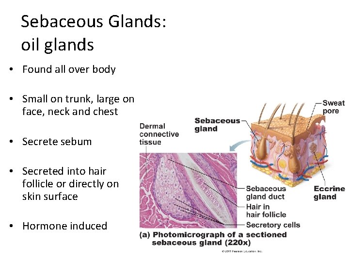
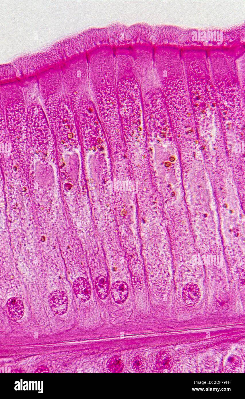
Label the photomicrograph of the skin and its accessory structures.. Solved Label the photomicrograph of the skin and its - Chegg See the answer Label the photomicrograph of the skin and its accessory structures Epidermis Duct of sebaceous gland Hair follicle Sebaceous gland Show transcribed image text Expert Answer 2. The picture here demonstrates the pseudostratified columnar epithelium. Figure 7.4 Photomicrograph of the skin and accessory structures - Quizlet Papilla of hair. a projection of connective tissue into the hair follicle and contains blood vessels that provide nutrients to the dividing cells of the matrix. Sebaceous Gland. Oil glands that surround hair follicles; secrete oils that lubricates skin, hair, and into the neck of the hair follicle. Hair Follicle. pg 87 fig 7.4 diagram of skin and accessory structures Start studying pg 87 fig 7.4 diagram of skin and accessory structures. Learn vocabulary, terms, and more with flashcards, games, and other study tools. Solved Label the photomicrograph of the skin and its | Chegg.com Label the photomicrograph of the skin and its accessory structures. Epidermis Sebaceous gland Hair follicle Duct of sebaceous gland Label the photomicrograph of the skin and its accessory structures. Epidermis Sebaceous gland Hair follicle Duct of sebaceous gland ; Question: Label the photomicrograph of the skin and its accessory structures ...
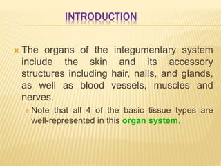
Structure and Function of Skin | Biology for Majors II - Course Hero The hypodermis (also called the subcutaneous layer or superficial fascia) is a layer directly below the dermis and serves to connect the skin to the underlying fascia (fibrous tissue) of the bones and muscles. It is not strictly a part of the skin, although the border between the hypodermis and dermis can be difficult to distinguish. Answered: 1. In the photomicrograph below of… | bartleby 1. In the photomicrograph below of cartilage tissue, find and label the indicated structures. Extra cellular r Lacuna Chondrocyte Dyte Elastic protein fibers Extracellular matrix In the photomicrograph below of compact bone tissue, find and label the indicated structu p Osteon Lamella Lacuna o Osteocyte Canaliculi Central canal Question Accessory Structures of the Skin | Anatomy and Physiology I - Course Hero Accessory structures of the skin include hair, nails, sweat glands, and sebaceous glands. These structures embryologically originate from the epidermis and can extend down through the dermis into the hypodermis. Hair Figure 1. Hair follicles originate in the epidermis and have many different parts. A&P 1 Exercise_7 Activity 1 & 2 & RYK and UYK.docx - LAB... Label the diagram of the skin and accessory structures in Figure 7.3. 1. Hair Shaft 2. Hair root 3. Sebaceous (oil) glands 4. Arrector Pilli Muscle 5. Hair follicle 6. Hair bulb 7. Eccrine sweat Gland 8. Papillae of hair 9. Apocrine sweat Gland Label the photomicrograph in Figure 7.4. 1. Sebaceous glands 2. Hair follicle 3. Hair root 4.
photomicrograph of thick skin Diagram | Quizlet Start studying photomicrograph of thick skin. Learn vocabulary, terms, and more with flashcards, games, and other study tools. Home. Subjects. Explanations. Create. Study sets, textbooks, questions. ... skin structure (part 1 of 2) 8 terms. mckennawebber. skin structure (part 2 of 2) 8 terms. mckennawebber. nail structure (part 1 of 2) 8 terms. Layers of the Skin | Anatomy and Physiology I - Lumen Learning The hypodermis (also called the subcutaneous layer or superficial fascia) is a layer directly below the dermis and serves to connect the skin to the underlying fascia (fibrous tissue) of the bones and muscles. It is not strictly a part of the skin, although the border between the hypodermis and dermis can be difficult to distinguish. Anatomy of the Epidermis with Pictures - Verywell Health The four layers of cells, beginning at the bottom, are the stratum basale, stratum spinosum, stratum granulosum, and stratum corneum. In your palms and soles, there's an additional layer called stratum lucidum underneath the stratum corneum. In the bottom layer, keratinocytes divide and push up formed cells toward the upper layer. PDF CHAPTER 5 The Integumentary System - LWW The skin consists of two layers: the epidermis, which includes the outer protective stratum of keratinized epithelial cells, and the dermis, the underlying layer of connective tissue containing blood vessels and nerve endings. The accessory structures include outgrowths of the epidermis (hair and nails).
Full article: Identification of Mammal Skins Present in an ...
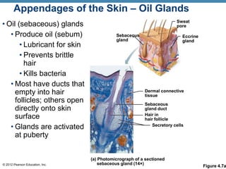
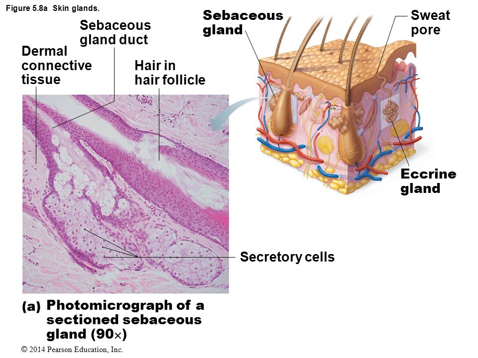
Accessory Structures of the Skin - Anatomy & Physiology Accessory structures of the skin include hair, nails, sweat glands, and sebaceous glands. These structures embryologically originate from the epidermis and can extend down through the dermis into the hypodermis. Hair Hair is a keratinous filament growing out of the epidermis. It is primarily made of dead, keratinized cells.
Solved Label the photomicrograph of the skin and its | Chegg.com
Figure 7.1: Photomicrograph of Skin Diagram | Quizlet Start studying Figure 7.1: Photomicrograph of Skin. Learn vocabulary, terms, and more with flashcards, games, and other study tools.

5.1 Layers of the Skin – Anatomy & Physiology
Diagram of human skin structure — Science Learning Hub Diagram of human skin structure. Image. Add to collection. Tweet. Rights: University of Waikato Published 1 February 2011 Size: 100 KB Referencing Hub media. The epidermis is a tough coating formed from overlapping layers of dead skin cells.

Integumentary system Module 3: Accessory Structures of the ...
Solved Label the Photomicrograph of the skin and its - Chegg Expert Answer. 100% (3 ratings) Transcribed image text: Label the Photomicrograph of the skin and its accessory structures.

anatomy lab, exam 3, lab 9, Spinal Nerves, Integument, and ...
Diagram of the skin and accessory structures - Quizlet Start studying Diagram of the skin and accessory structures. Learn vocabulary, terms, and more with flashcards, games, and other study tools.
Integumentary System 2 Regions 1 Epidermis 2 Dermis
Skin Anatomy: The Layers of Skin and Their Functions - Verywell Health Subcutaneous tissue is the innermost layer of the skin. It is mostly made up of fat, connective tissues, larger blood vessels, and nerves. 5 The majority of your body fat is stored in the subcutaneous layer. It not only insulates you against changing temperatures but protects your muscles and internal organs from impacts and falls.

SciELO - Brasil - Macroscopic and microscopic morphology of ...
PDF Name the Condition - Dr. Scott Croes' Website the cartoon and the photomicrograph. •Name the Layers of skin and label the dermal papilla and dermis •Name the Layers of skin and label the dermal papilla and dermis. Name the layer of skin shown. Stratum Spinosum. Name the specific layers of skin indicated by the ... Identify the following structures: Epidermis, Hair cortex, Hair medulla ...

A&P Chapter 4 - Diagram of the Skin Flashcards | Quizlet
unit 4 lab.docx - LAB Unit 4 EXERCISE 7: The Integumentary... FIGURE 7.4:Diagram of the skin and accessory structures. • apocrine (AP-oh-krin) sweat gland • arrector pili (PIE-lee) muscle • eccrine (EK-rin) sweat gland • hair bulb • hair follicle • hair root • hair shaft • papilla (puh-PILL-uh) of hair • sebaceous (se-BAY-shus) gland 1. Hair shaft 2. Hair root 3. Sebaceous glands 4. Arrector pili muscle 5.

Finger longitudinal section hi-res stock photography and ...
Integumentary System HW_answers.docx - Course Hero 18.Label the photomicrograph of the skin and its accessory structures. 19.Label the photomicrograph of the sebaceous gland. 20.Label the structures of merocrine sweat glands. 21.Label the structures of the hair follicle.

Skin 2: accessory structures of the skin and their functions ...
Skin 2: accessory structures of the skin and their functions All are important in the skin's key functions, including protection, thermoregulation and its sensory roles. This article, the second in a two-part series, looks at the structure and function of the main accessory structures of the skin. Citation: Lawton S (2020) Skin 2: accessory structures of the skin and their functions.
Integumentary System Overview
Solved Label the photomicrograph of the skin and its - Chegg Question: Label the photomicrograph of the skin and its accessory structures. Sebaceous gland Duct of sebaceous gland Epidermis Hair follicle This problem has been solved! See the answer Show transcribed image text Expert Answer 100% (19 ratings)
The Integumentary System
PDF Integumentary Answers - Home - Holly H. Nash-Rule, PhD Integumentary Answers - Home - Holly H. Nash-Rule, PhD
Hautquerschnitt -Fotos und -Bildmaterial in hoher Auflösung ...
Label the photomicrograph in Figure 7.4. Examine a slide of hairy skin ... Label the layers of the skin on the diagram and the photograph. Be able to identify the layers on a microscope slide. Look at the skin slide under a microscope. a) Epidermis 1) Stratum corneum ii) Stratum lucidum 111) Stratum granulosum iv) Stratum... Posted one year ago Recent Questions in Basics of Statistics Q:

SciELO - Brasil - Macroscopic and microscopic morphology of ...

unit 4 lab.docx - LAB Unit 4 EXERCISE 7: The Integumentary ...
Handout: The Integumentary System Anatomy & Physiology I ...

Hair, Hair Follicles, Sebaceous Glands, and Sudoriferous ...

Layers of the Skin | Anatomy and Physiology I
Chapter 5
Integumentary System Overview
Solved Label the photomicrograph of the skin and its | Chegg.com

Anatomy of the Lymphatic and Immune Systems | Anatomy and ...

5.1 Layers of the Skin – Anatomy & Physiology

glandular hairs - Keyword Search - Science Photo Library

PPT - Integumentary System PowerPoint Presentation, free ...

Transverse sections of the external genitalia of male and ...
Simple ciliated columnar hi-res stock photography and images ...
Ch 4 Skin Power Point
5 The Integumentary System Pages , ppt video online download

Lap Practical #1 EC Flashcards | Quizlet

unit 4 lab.docx - LAB Unit 4 EXERCISE 7: The Integumentary ...
A Comparative Account of the Morphological and Anatomical ...
The Integumentary System

5.1 Layers of the Skin – Anatomy & Physiology
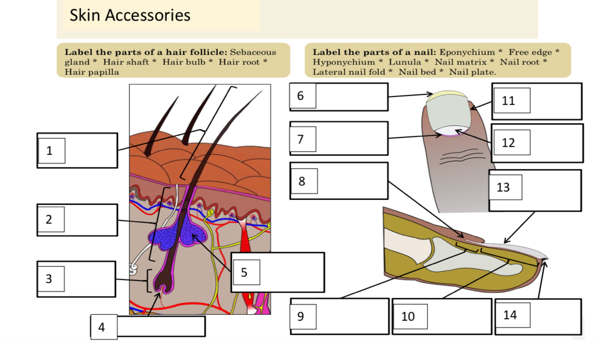
Answered: Label the parts of a hair follicle:… | bartleby
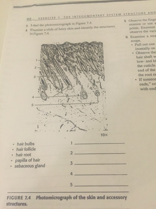
Solved Label the photomicrograph in Figure 7.4. Examine a ...

Skin Structure (Labeling) Flashcards | Quizlet
PPT - Integumentary System PowerPoint Presentation, free ...
Dentin-pulp complex development – Histology and Embryology ...
Integumentary System Overview
Novel quantitative signature of tumor stromal architecture ...
The Respiratory System
Post a Comment for "42 label the photomicrograph of the skin and its accessory structures."