44 sheep brain diagram labeled
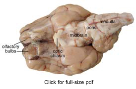
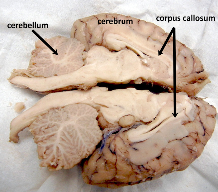
Sheep Brain Dissection Project Guide | HST Learning Center Sheep Brain Dissection: Internal Anatomy. Place the brain with the curved top side of the cerebrum facing up. Use a scalpel (or sharp, thin knife) to slice through the brain along the center line, starting at the cerebrum and going down through the cerebellum, spinal cord, medulla, and pons. Separate the two halves of the brain and lay them ... PDF Sheep Brain Midsagittal Section - Dr. Scott Croes' Website Sheep Brain -Parasagittal Section 1. Gray Matter 2. White Matter 3. Corpus Callosum 4. Lateral Ventricle 5. Caudate Nucleus 6. Septum Pellucidum 7. Fornix 8. Optic Chiasma 9. Third Ventricle 10. Thalamus (Ovid Nuclear Mass of Thalamus) 11. Corona Radiata 12. Hippocampus 13. Cerebral Aqueduct (of Sylvius) 14. Pituitary Gland (hypophysis) 15.
labeled diagram of the brain Midsagittal Sheep Brain (labeled) | anatomyphyslab261 | Flickr we have 8 Pics about 25. Midsagittal Sheep Brain (labeled) | anatomyphyslab261 | Flickr like Brain And Spinal Cord Diagram Anatomy Chart Of Spinal Cord Labeled, 25. ... brain diagram unlabeled human half anatomy clipart left clip blank cliparts labeled psych ap results etc ...
Sheep brain diagram labeled
Solved Art-labeling Activity: Midsagittal Section of the - Chegg Anatomy and Physiology. Anatomy and Physiology questions and answers. Art-labeling Activity: Midsagittal Section of the Sheep Brain (Diagram, 2 of 2) Reset Help Fomix Infundibulum Olfactory bulb Optic chiasm Mosencephalon Pituitary gland Marillary body Medulla oblongata Pons Spinal cord Corpus callosum Art-labeling Activity: Midsagittal Section ... Sheep Brain Label - sheep brain 2, 34 label the sheep brain labels ... Sheep Brain Label - 16 images - label the parts of a sheep brain midsagittal, superior view of sheep s brain, brain corpus callosum and ventricles, pretty good picture of the sheep brain labeled brain anatomy basic, brain diagram unlabeled Spinal cord anatomy nerves nervous quiz system brain mater autonomic commissure gray nerve exercise quizlet boundary anterior dissection sheep superior Pig Heart Diagram Labeled : Biological Science Picture Directory. 9 Pictures about Pig Heart Diagram Labeled : Biological Science Picture Directory : Pin on unlabeled Anatomy, Midsagittal ...
Sheep brain diagram labeled. label the brain anatomy diagram Pixelated Brain: Neuroanatomy For Medical Students brain anatomy neuroscience answer Brain sheep label anatomy labeled superior classroom labels sdmesa physiology nervous inferior edu lateral sup ventricle bottom section sagittal creative. Nervous system worksheet answers. Sheep brain dissection lab companion Label Sheep Brain Diagram | Quizlet Label Sheep Brain STUDY Learn Flashcards Write Spell Test PLAY Match Gravity + − Created by telaneyn Terms in this set (11) Cerebellum the "little brain" at the rear of the brainstem; functions include processing sensory input and coordinating movement output and balance Occipital lobe Sheep Brain Images - San Diego Mesa College Sheep Brain Unlabeled. Sheep Brain Leader-Lined. Sheep Brain Labeled. San Diego Mesa College 7250 Mesa College Drive San Diego, CA 92111-4998 Student Support San Diego Community College District San Diego City College San Diego Mesa College San Diego Miramar College San Diego Continuing Education. DOC Sheep Brain Anatomy Lab Manual - amherst.edu Sheep Brain Anatomy Lab Manual. Based on original material by R. N. Leaton, Dartmouth College. Contributors to this version: Al Sorenson, Lisa Raskin, Sarah Turgeon, Steve George, and JP Baird. I. Introduction. The brain of the sheep is useful for study because its anatomy is similar to human brain anatomy. Although exact proportions (and names ...
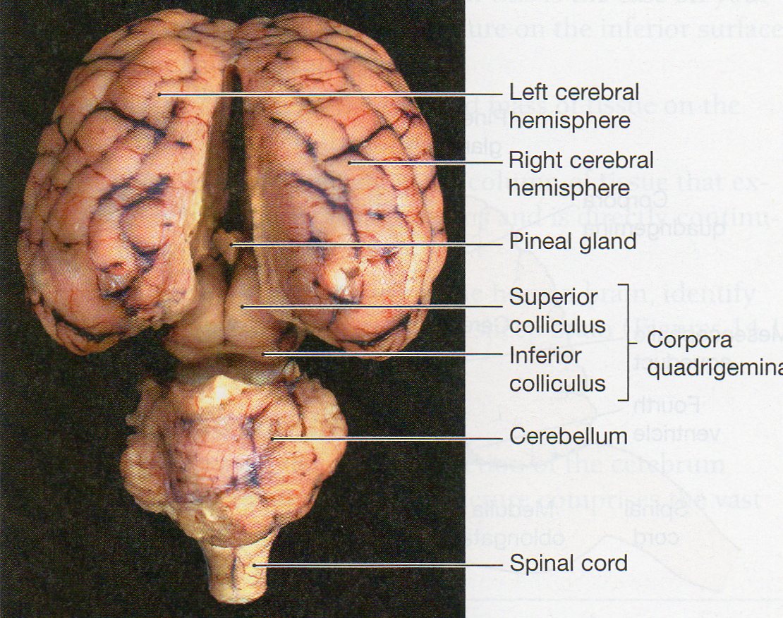
Sheep Brain Dissection labeled Diagram | Quizlet Only $2.99/month Sheep Brain Dissection labeled STUDY Learn Write Test PLAY Match Created by AllieKlinger Terms in this set (8) Corpus Collosum Lateral Ventricle Fornix Hypothalamus Cerebral Aqueduct Central Canal Inferior Collicuious Transverse Fissure THIS SET IS OFTEN IN FOLDERS WITH... Sheep Brain Dissection labeled 2 8 terms AllieKlinger brain parts labelled diagram brain sheep label anatomy labeled superior classroom labels sdmesa physiology nervous inferior edu lateral sup ventricle section sagittal creative coronal Brain anatomy function lobe sagittal lateral frontal occipital normal medicalartworks temporal parietal anatomical psychology neuroscience. Dog eye anatomy. Brain stem and adjacent structures sheep anatomy diagram Dr. Parker's A&P I Sheep Brain Dissection - YouTube we have 17 Images about Dr. Parker's A&P I Sheep Brain Dissection - YouTube like Pin by Kenton Antiques on Vintage Antique Ephemera | Sheep, Ewe sheep, Ovine and Caprine Husbandry | Veterian Key | Sheep, Animal anatomy, Anatomy and also Biology 2404. ... Ewe Sheep . sheep ... labeled diagrams of the brain labeled diagrams of the brain Brain corpus sheep callosum ventricle ventricles fornix third dissection labeled thalamus between space fluid lateral filled main structures taken been. Sheep brain dissection nervous system activity science teacherspayteachers. Related image labeled diagrams of the brain
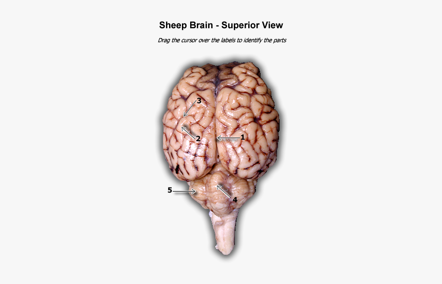
brain diagram nervous system anatomy human brain anatomy - Sheep Brain ... Read Or Download Gallery of brain diagram nervous system anatomy human brain anatomy - Sheep Brain Label | anatomical brain stem labeled lab model google search anatomy, sheep brain dissection bi biology junction, 31 label the sheep brain, 34 label the sheep brain labels design ideas 2020, Sheep Brain Anatomy Quiz - ProProfs 1. What is the outer covering of the Sheep brain? A. Arachnoid Mater B. Pia Mater C. Medulla Oblongata D. Dura Mater 2. Which amongst the following is the largest structure in dorsal view? A. Pons B. Medulla Oblongata C. Cerebral cortex D. Hippocampus 3. What are the large folds that surround the cerebrum? A. Gyri B. Sulci C. Fissures D. Colliculus the brain diagram labeled sheep brain dissection parker dr Pin On Unlabeled Anatomy nervous system unlabeled Human Circulatory System Diagram Labeled Circulatory System The Free heart diagram human circulatory labeled system simple anatomy circulation attack notes Click On The Links Below To See Labeled Images Of A Dissected Pigeon Practice Lab Practical on the Sheep Brain Identify the lobe labeled 1. Identify the lobe labeled 2. Identify the lobe labeled 3. Identify the structure labeled 4. Identify the structure lobe labeled 5. ... Identify the shiny membrane visible on the sheep brain surface. In the above picture: Identify the structure labeled 1. Identify the structure labeled 2. ...
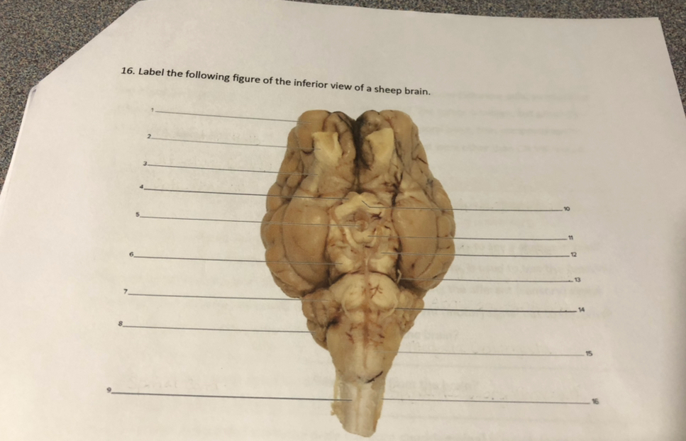
Solved 1. Label the following figure of the inferior view of ...
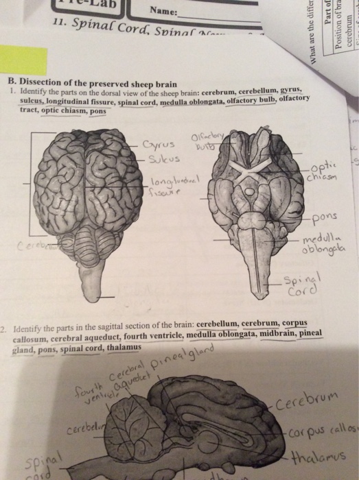
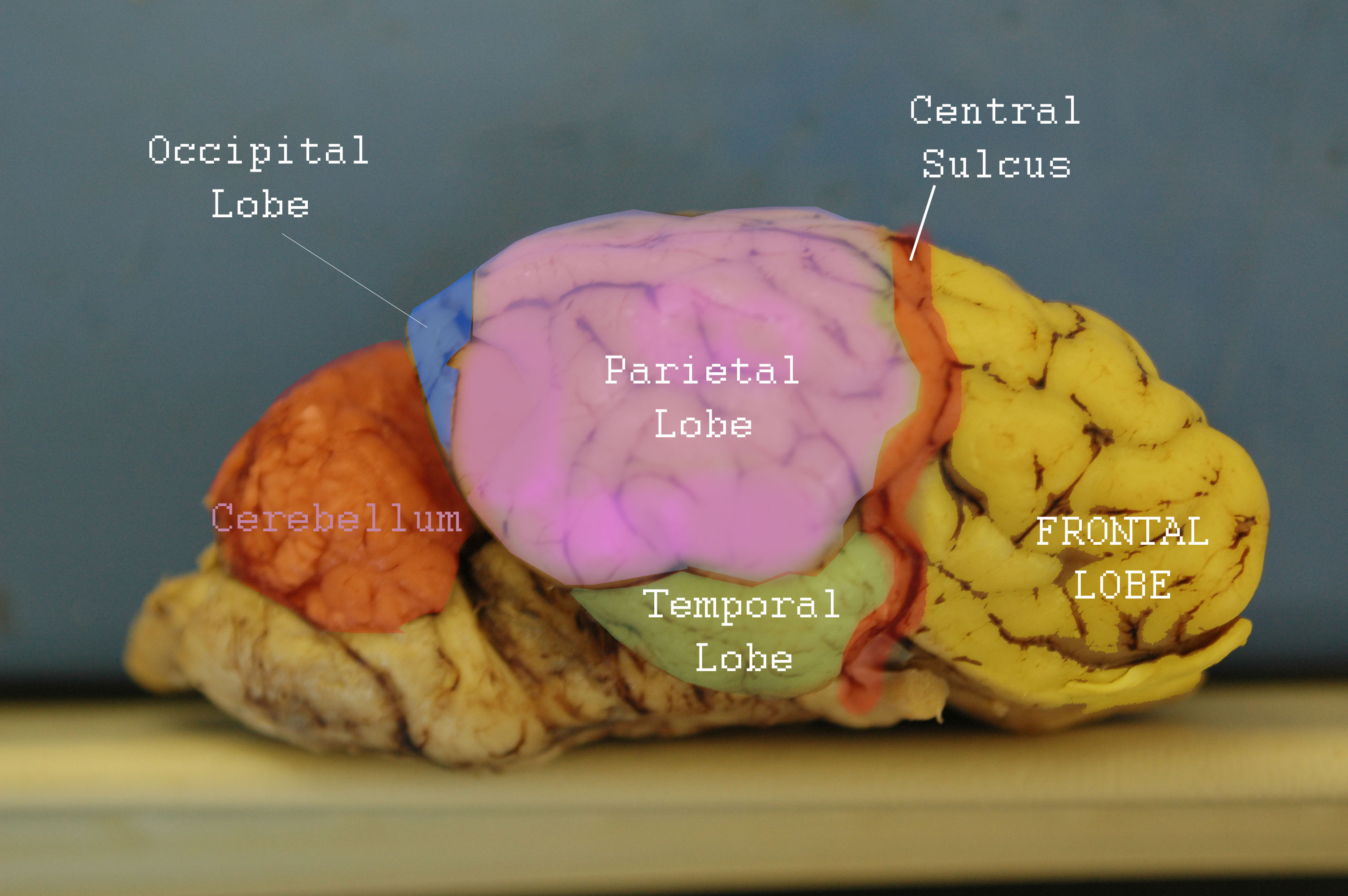
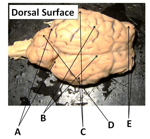
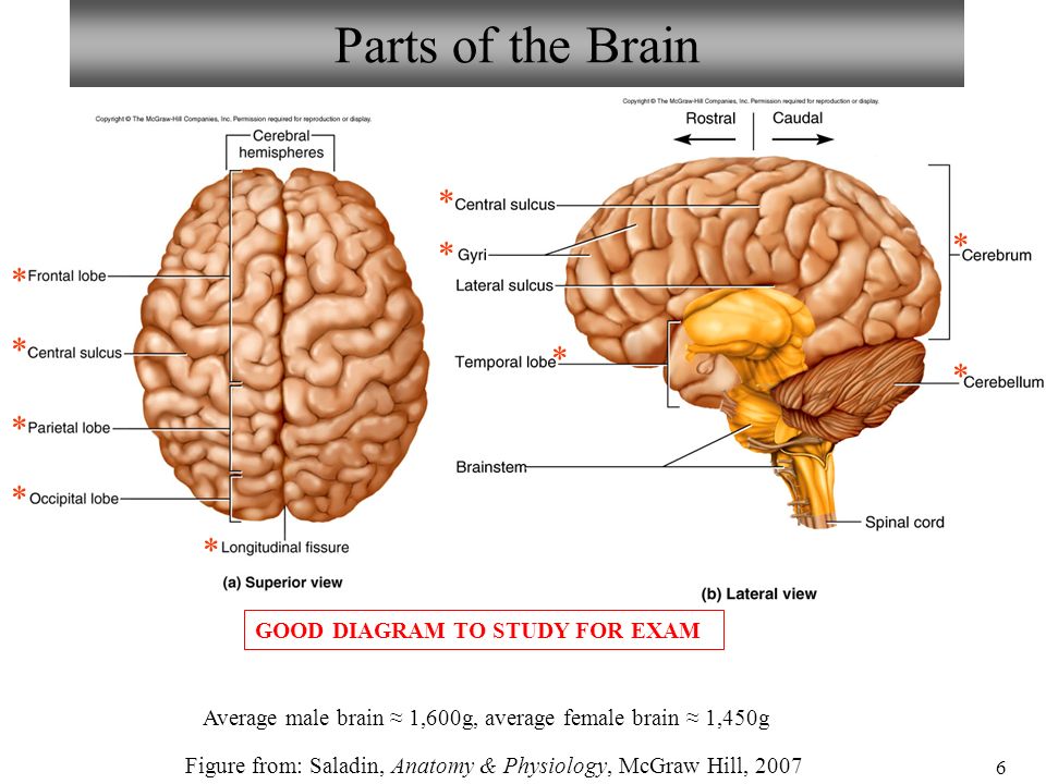
PDF Sheep Neuroanatomy Lab- Labeling Worksheet Psychology 2315- Brain and ... Sheep Neuroanatomy Lab- Labeling Worksheet Psychology 2315- Brain and Behaviour Kwantlen Polytechnic University Figure 1: Dorsal view Cerebellum, Frontal lobe, Occipital lobe, Parietal lobe, and Temporal lobe. Temporal Parietal Lobe Frontal Lobe Cerebellum Occipital Lobe

ImageQuiz: Sheep brain dorsal view
labeled brain | Brain anatomy, Anatomy and physiology, Anatomy labeled brain | Brain anatomy, Anatomy and physiology, Anatomy From anatomycorner.com Sheep Brain Images taken from the dissection of the sheep's brain: cerebrum, cerebellum, corpus callosum, lobes, sulci, gyrus, fornix, pituitary. H Hillary Hunt 322 followers More information labeled brain Find this Pin and more on NeuroBiology by Hillary Hunt.
Sheep brain dissection - Bisc 163 - StuDocu
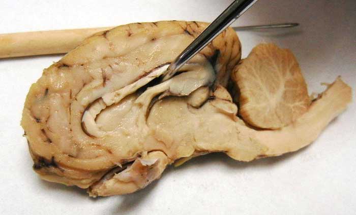
PDF Neuroanatomy: Dissection of the Sheep Brain - Napa Valley College Examine the sheep brain with the membranes intact. You should be able to identify and use the following directional terms: Anterior / Posteriorfront / back Rostral / Caudal towards the beak / towards the tail Medial / Lateral towards the middle / towards the side Dorsal / Ventral top / bottom (on the CNS of a quadruped)
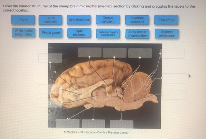
Solved Label the interior structures of the sheep brain ...
Sheep Brain Label | Dissection, Human brain diagram, Brain anatomy Sheep Brain Label A drawing of the brain with the parts unlabeled. Students can practice naming the parts of the brain, then check their answers with the provided key. Biologycorner 17k followers More information unlabeled brain Find this Pin and more on A&P by Dijana Kovacevic. Human Brain Diagram Brain Gym For Kids Brain Anatomy And Function
Neuroanatomy: Dissection of the Sheep Brain
brain diagram anatomy Vintage Anatomy Skull Image - The Graphics Fairy thegraphicsfairy.com. skull anatomy fairy. 027 The 3 Parts Of The Brain Stem And Their Functions - YouTube . brain stem parts functions vzo. Brain label anatomy cranial nerves labeled labels models human inferior somso nervous base gross. Schwann cells keep signals strong.

Sheep Brain Dissection
nervous system diagram labeled brain Sheep Brain Dissection Lab Companion jb004.k12.sd.us sheep brain dissection mater dura anatomy lab layers superior dorsal diagrams cranial use companion bi nervous system names visit quizlet Artery And Vein Model | Arteries And Veins, Anatomy Models, Arteries

Sheep Brain: Top View Diagram | Quizlet
Sheep Brain Dissection | Carolina.com Jun 6, 2017 — Carolina's Perfect Solution® sheep brain dissection introduces students to the anatomy of a mammalian brain, both external and internal, and ...

Sheep Brain Dissection | Carolina.com
PDF DISSECTION OF THE SHEEP'S BRAIN - Hanover College DISSECTION OF THE SHEEP'S BRAIN Introduction The purpose of the sheep brain dissection is to familiarize you with the three-dimensional structure of the brain and teach you one of the great methods of studying the brain: looking at its structure. One of the great truths of studying biology is the saying that "anatomy precedes physiology".

Sheep brain | Atlas of Comparative Vertebrate Anatomy
brain anatomy cerebral cortex Sheep brain anatomy label nervous system dissection lateral ventricle section sagittal sdmesa classroom edu cerebral diagram aqueduct lesson nerves cranial. Brain area google edu csus cortex areas primary cortical auditory wernicke result indiv forebrain premotor доску выбрать areas2. Frontal Lobes - Motor Cortex, Cognition, and ...
Lab Exam 3: Anatomy of Sheep Brain; Histology Flashcards ...
Sheep Brain Dissection with Labeled Images 1. The sheep brain is enclosed in a tough outer covering called the dura mater. You can still see some structures on the brain before you remove the dura mater. Take special note of the pituitary gland and the optic chiasma. These two structures will likely be pulled off when you remove the dura mater. Brain with Dura Mater Intact

Resources for Teaching Mammalian Neuroanatomy Using Sheep ...
sheep anatomy diagram sagittal sheep brain spinal cord mccc edu Sheep Brain #2 sheep brain anatomy label nervous system dissection lateral ventricle section sagittal sdmesa classroom edu cerebral diagram aqueduct lesson nerves cranial Anatomy Of A Sheep suffolksheepni.co.uk sheep anatomy external artist names Sheep Brain Flashcards | Quizlet

Pin page
Sheep brain dissection | Human Anatomy and Physiology Lab (BSB 141 ... The sheep brain is quite similar to the human brain except for proportion. The sheep has a smaller cerebrum. Also, the sheep brain is oriented anterior to posterior (more horizontally), while the human brain is oriented superior to interior (more vertically.) ... 91400 Human Anatomy and Physiology - Practical Manual Summer 2021 - Part 1 (1).pdf.

SHEEP BRAIN - Biology Forums Gallery
brain diagram unlabeled Spinal cord anatomy nerves nervous quiz system brain mater autonomic commissure gray nerve exercise quizlet boundary anterior dissection sheep superior Pig Heart Diagram Labeled : Biological Science Picture Directory. 9 Pictures about Pig Heart Diagram Labeled : Biological Science Picture Directory : Pin on unlabeled Anatomy, Midsagittal ...
Solved 1. Identify the parts on the dorsal view of the sheep ...
Sheep Brain Label - sheep brain 2, 34 label the sheep brain labels ... Sheep Brain Label - 16 images - label the parts of a sheep brain midsagittal, superior view of sheep s brain, brain corpus callosum and ventricles, pretty good picture of the sheep brain labeled brain anatomy basic,
Sheep Brain Dissection with Labeled Images
Solved Art-labeling Activity: Midsagittal Section of the - Chegg Anatomy and Physiology. Anatomy and Physiology questions and answers. Art-labeling Activity: Midsagittal Section of the Sheep Brain (Diagram, 2 of 2) Reset Help Fomix Infundibulum Olfactory bulb Optic chiasm Mosencephalon Pituitary gland Marillary body Medulla oblongata Pons Spinal cord Corpus callosum Art-labeling Activity: Midsagittal Section ...

Sheep Brain Images
Labeled Superior View Of Real Brain, HD Png Download - kindpng
Physiological Psychology
Index of /files/OCC_VIDEO/upload/Faculty_Resources/acamilo ...
Lab: Sheep Brain Dissection
Sheep Brain Dissection Project Guide | HST Learning Center

sheep brain Diagram | Quizlet

Sheep Brain Dissection – Krysta H, Rinda G, Keira N, Cindy M ...

Sheep Brain Images

Sheep brain images | Lab | Amherst College

Sheep Brain Images
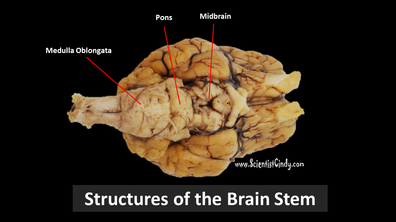
The Brain - SCIENTIST CINDY
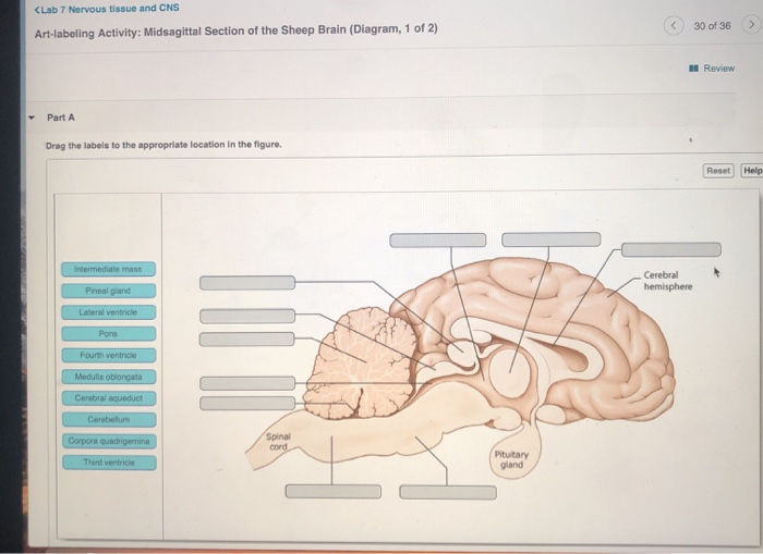
Solved art-labeing activity: midsagittao section of the ...
Sheep Brain Dissection with Labeled Images

Sheep brain | Atlas of Comparative Vertebrate Anatomy
Sheep Brain Dissection with Labeled Images

BIO201-Sheep Brain
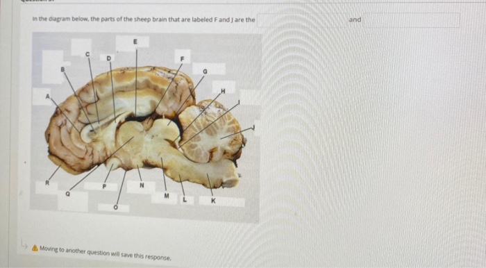
Solved in the diagram below, the parts of the sheep brain ...
Brain Anatomy Labeled Diagram Stock Vector - Illustration of ...
Medical Detectives Lesson 28

Sheep Brain
Lab - Sheep Brain: MAH-Summer 2019-Anatomy and Physiology I

Sheep Brain Images

Sheep Brain Diagram
Sheep Brain Neuroanatomy Online Self-Test | KPU.ca - Kwantlen ...
Lab: Sheep Brain Dissection
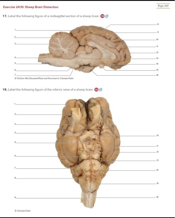
Solved Exercise 24.10: Sheep Brain Dissection Page 509 ...
Sheep Neuroanatomy Lab- Labeling Worksheet Figure 1: Dorsal view
Neuron/Spinal Cord Histology Brain Anatomy Sheep Brain ...
Post a Comment for "44 sheep brain diagram labeled"