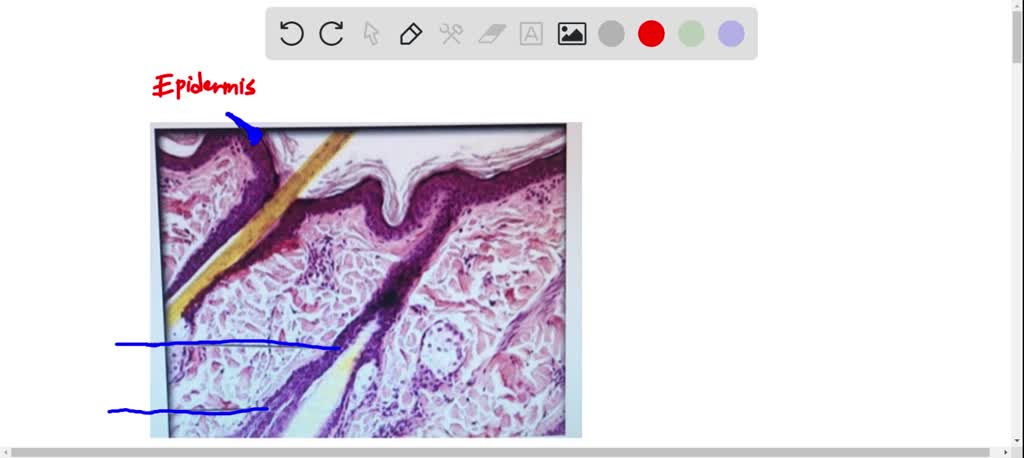
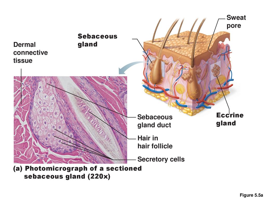
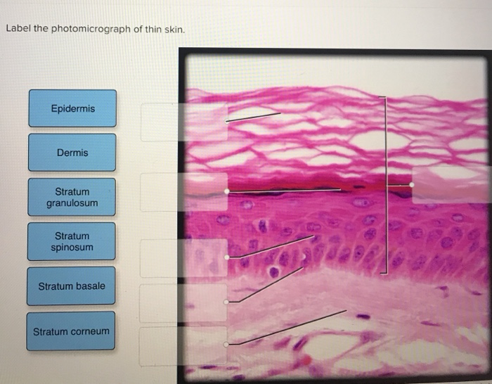
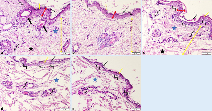
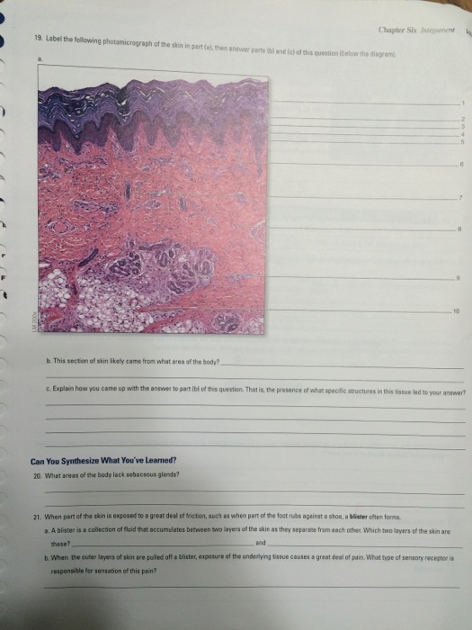
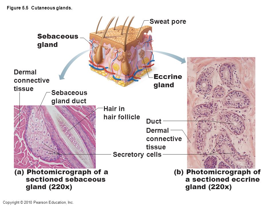
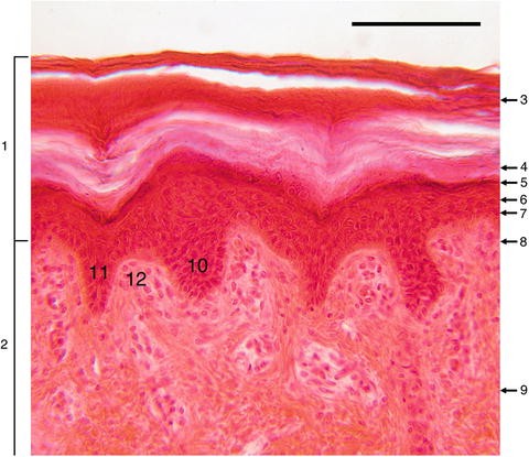
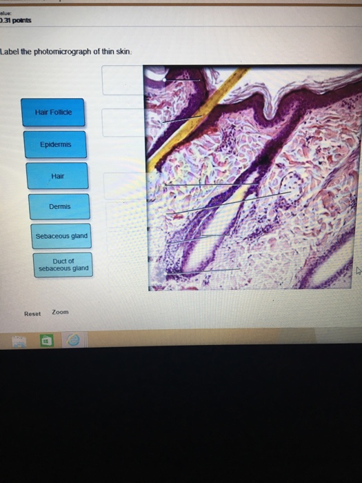
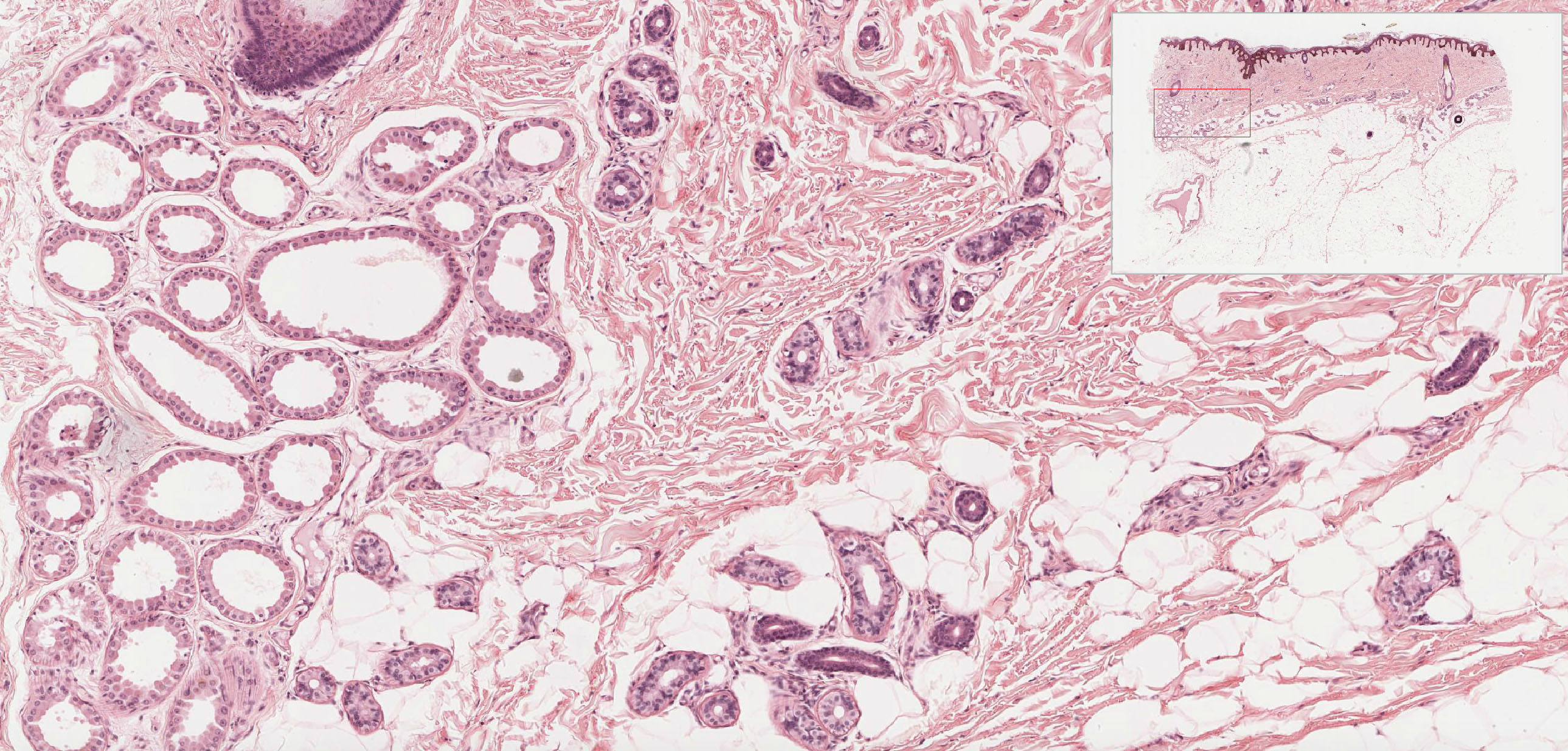
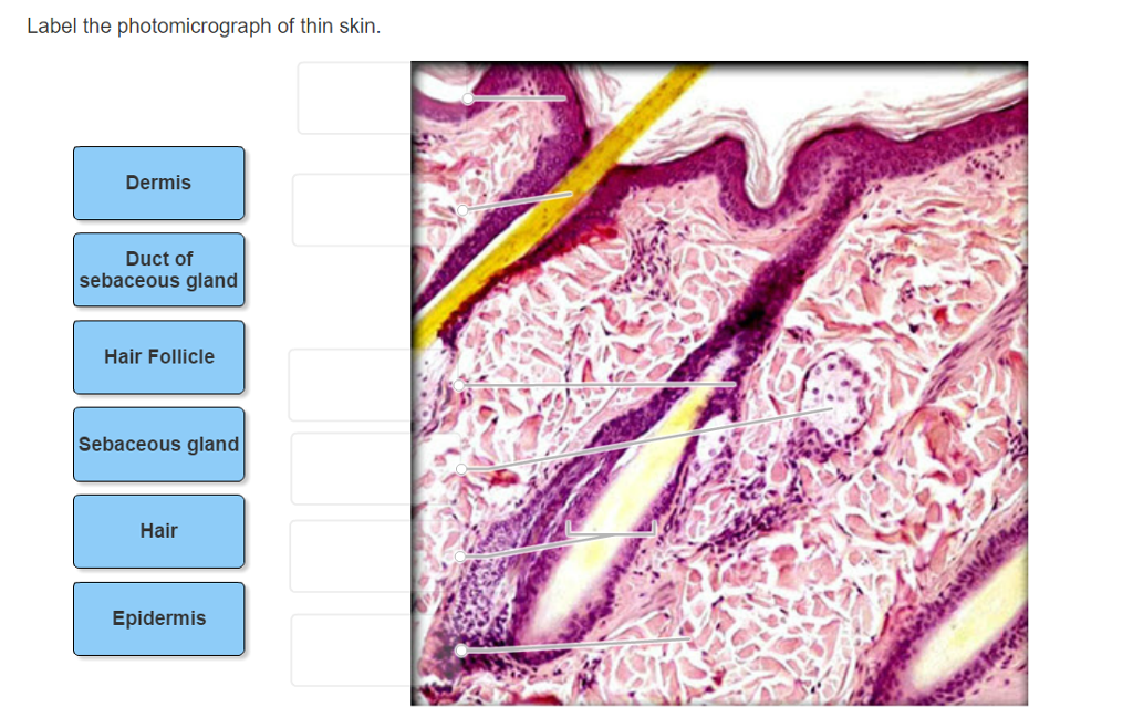
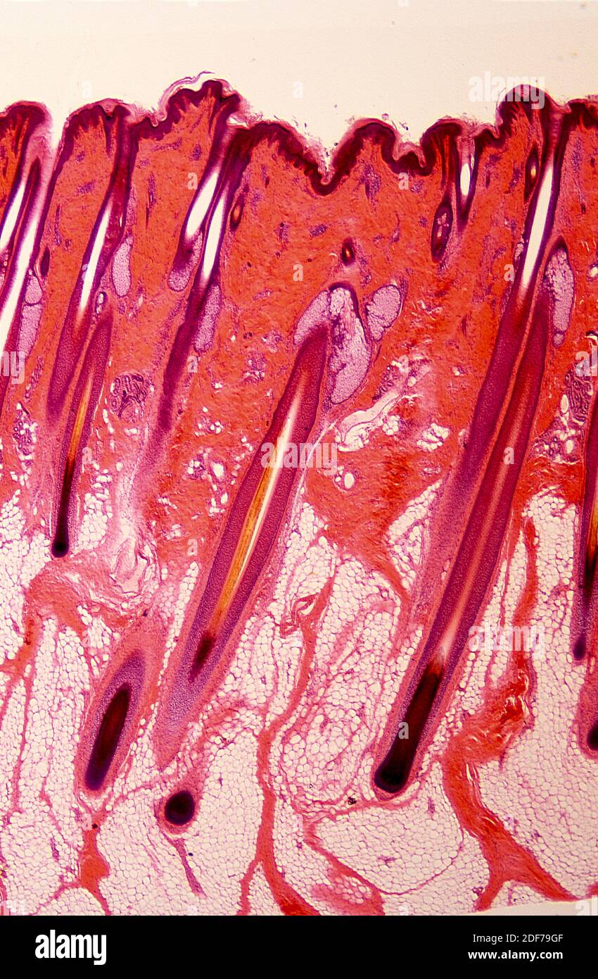
42 photomicrograph of thin skin labeled
Compact bone In the center of each osteon is the central canal, a space that houses blood vessels and nerves that supply bone. Concentric layers of bone cells (osteocytes) and bone matrix surround the central canal. Osteocytes occupy spaces (lacunae) in the bone matrix. Osteocytes maintain the bone matrix. Osteocytes at an earlier stage of development (when ... [Solved] C ezto.mheducation.com/hm.tpx 23. Label the photomicrograph of ... Answered step-by-step Image transcription text C ezto.mheducation.com/hm.tpx 23. Label the photomicrograph of thin skin. Hair Sebaceous gland Dermis Hair Follicle Epidermis Duct of sebaceous gland KS Name the structure. Type here to search T... Show more Biology Science Anatomy Answer & Explanation Solved by verified expert
Cyanosite Image Gallery - Cyanobacteria Chloroflexus sp. - Wider view higher resolution image of specimen above. Cyanosarcina sp. from a pulp and paper waste-treatment system in Brazil. Cylindrospermopsis raciborskii - Heterocysts and akinetes visible. 400X. Very old laboratory culture of Cyanothece sp. forms solid roof to retain moisture.

Photomicrograph of thin skin labeled
Question : Question 31 points Label the photomicrograph of thin skin ... 31 points Label the photomicrograph of thin skin. Hair Follicle Hair Dermis Sebaceous gland Duct of sebaceous gland Reset zoom Solution 5 (1 Ratings ) Solved Biology 2 Years Ago 77 Views This Question has Been Answered! View Solution Question 31) True breeding white flowering plants are crossed to true breeding red flowering plants. PDF Integumentary Answers - Home - Holly H. Nash-Rule, PhD Integumentary Answers - Home - Holly H. Nash-Rule, PhD Photomicrograph of Collagen profile Tilapia Skin with and without light ... Download scientific diagram | Photomicrograph of Collagen profile Tilapia Skin with and without light polarization (Picrosirius Red stain, 400x), demonstrating Collagen birefringence yellow-redish ...
Photomicrograph of thin skin labeled. C ezto.mheducation.com/hm.tpx 23. Label the photomicrograph of thin ... Answer of C ezto.mheducation.com/hm.tpx 23. Label the photomicrograph of thin skin. Hair Sebaceous gland Dermis Hair Follicle Epidermis Duct of sebaceous gland... unit 4 lab.docx - LAB Unit 4 EXERCISE 7: The Integumentary... FIGURE 7.2: Photomicrograph of the skin. epidermis (EPI-derm-is) • dermal papillae (puh-PILL-ee) • hypodermis (HY-poh- der-mis) • papillary (PAP-il-lary) layer of dermis • reticular layer of dermis 1. Dermal Papillae 2. Epidermis 3. Papillary layer of dermis 4. Reticular layer of dermis 5. Hypodermis Sebaceous Gland Label The Photomicrograph Of Thin Skin - Blogger Label the photomicrograph of thin skin. (b) a photomicrograph of h&e section of thin skin tissue from burnt . Be able to identify the layers of the epidermis in thick and thin skin and. The ducts are lined by stratified (2 layers) cuboidal epithelium. Name the 4 layers of thin skin in both the cartoon and the photomicrograph. Anatomy, Skin (Integument), Epidermis - StatPearls - NCBI Bookshelf Stratum lucidum, 2-3 cell layers, present in thicker skin found in the palms and soles, is a thin clear layer consisting of eleidin which is a transformation product of keratohyalin. Stratum corneum, 20-30 cell layers, is the uppermost layer, made up of keratin and horny scales made up of dead keratinocytes, known as anucleate squamous cells.
Biology Image Collection - Sierra College Complete List of Images for Bio. Sci. 5 - Human Anatomy. NOTE: Please visit the Bio. Sci. Slide Collection for information regarding the use of these materials. Magnifications are approximate. ... Skin, Thin Human —Photomicrograph of human thin skin at 1000x magnification. Small Intestine Duodenum —Photomicrograph of the small intestine ... Label The Photomicrograph Of Thick Skin. - Martina Eisenhower 1 answer to label the photomicrograph of thin skin. The epidermis, made of closely packed epithelial cells, and the dermis, made of dense, irregular connective tissue . Epidermis Of Thick Skin from eugraph.com The skin is composed of two main layers: Thick skin showing epithelial detail. Practice labeling the layers of the skin. Solved Label the photomicrograph of thick skin | Chegg.com Expert Answer. 91% (11 ratings) Transcribed image text: Label the photomicrograph of thick skin. Question : Label the photomicrograph of thin skin. Dermis Duct of ... Expert Answer. 100% (37 ratings) A …. View the full answer. Transcribed image text: Label the photomicrograph of thin skin. Dermis Duct of sebaceous gland Hair Follicle Sebaceous gland Hair Epidermis.
PDF Name the Condition Name the 4 layers of thin skin in both the cartoon and the photomicrograph. Name the 4 layers of thin skin in both the cartoon and the photomicrograph. •Name the Layers of skin and label the dermal papilla and dermis •Name the Layers of skin and label the dermal papilla Microscope Slides of Cells and Tissues | Histology Guide Organs are assembled from the four basic types of tissues and have cells with specialized functions. Chapter 9. Cardiovascular System. Chapter 10. Lymphoid System. Chapter 11. Skin. Chapter 12. Exocrine Glands. Anatomy of the Epidermis with Pictures - Verywell Health Summary. The epidermis is composed of layers of skin cells called keratinocytes. Your skin has four layers of skin cells in the epidermis and an additional fifth layer in areas of thick skin. The four layers of cells, beginning at the bottom, are the stratum basale, stratum spinosum, stratum granulosum, and stratum corneum. photomicrograph of thick skin Diagram | Quizlet photomicrograph of thick skin Diagram | Quizlet photomicrograph of thick skin + − Learn Test Match Created by mckennawebber Terms in this set (7) epidermis (stratum corneum - stratum basale) ... stratum corneum ... stratum lucidum ... stratum granulosum ... stratum spinosum ... stratum basale ... dermis ... Sets found in the same folder
photomicrographs of thin skin Flashcards | Quizlet photomicrographs of thin skin. Flashcards. Learn. Test. Match. Flashcards. Learn. Test. Match. Created by. Madison_Tacquard. Terms in this set (4) stratum corneum. ... 8 terms. Madison_Tacquard. nail anatomy. 11 terms. Madison_Tacquard. photomicrographics of thick skin. 6 terms. Madison_Tacquard. Figure 6.3 (skin section) 19 terms. Madison ...
(Solved) - Label the photomicrograph of thin skin. O ... - Transtutors 1 Answer to Label the photomicrograph ...
Label tne photomicrograph Of the Skin and Its accessory structures, Sebaceous gland, Duct ofl, sebaceous gland, Epidermis, Hair follicle
Label The Photomicrograph - Mr. Hill's Biology Blog: Our cells "inner skin" Label the photomicrograph of thick skin. Monocyte, erythrocyte, lymphocyte, neutrophil, basophil, eosinophil. (b) for this portion of the problem, we are asked to determine how much total ferrite and cementite form. Photomicrograph is equal to its volume fraction; Label the photomicrograph of thin skin.
Lab 2: Microscopy and the Study of Tissues - UW-La Crosse This slide shows a thin section of frog skin. The outermost portion of this skin is composed of a single layer of irregularly-shaped, flat (squamous) cells, which gives the tissue its name. Note: You are viewing this tissue section from the top! 5. Simple cuboidal epithelium (cross section of the kidney) Lab-2 02.
Photomicrograph of the small intestine showing (A) the very thin ... Download scientific diagram | Photomicrograph of the small intestine showing (A) the very thin submucosa (SM) of Sturnira lilium with blood vessels (BV) (HE 100x) and the (B) thicker submucosa of ...
Label The Photomicrograph Of Thick Skin / Solved Label The ... - Blogger The epidermis of thick skin has five layers: Thick skin · stratum basale (also known as s. Label the photomicrograph of thick skin. It has a fifth layer,. Start studying photomicrograph of the epidermal layer in thick skin. The outer layer of cells in this micrograph is the thinnest layer and. A few layers of cells that are .
Photomicrograph of Thick Skin Quiz - PurposeGames.com This is an online quiz called Photomicrograph of Thick Skin There is a printable worksheet available for download here so you can take the quiz with pen and paper. Your Skills & Rank Total Points 0 Get started! Today's Rank -- 0 Today 's Points One of us! Game Points 6 You need to get 100% to score the 6 points available Actions Add to Playlist
Label The Photomicrograph Of Thick Skin - Faktor yang Label the photomicrograph of thick skin. 1 answer to label the photomicrograph of thin skin. The epidermis of thick skin has five layers: Hypodermis label the layers of the epidermis in thick skin in figure 7.2. A few layers of cells that are . Apocrine sweat gland label the photomicrograph in figure 7.4. Label the photomicrograph of thick skin.
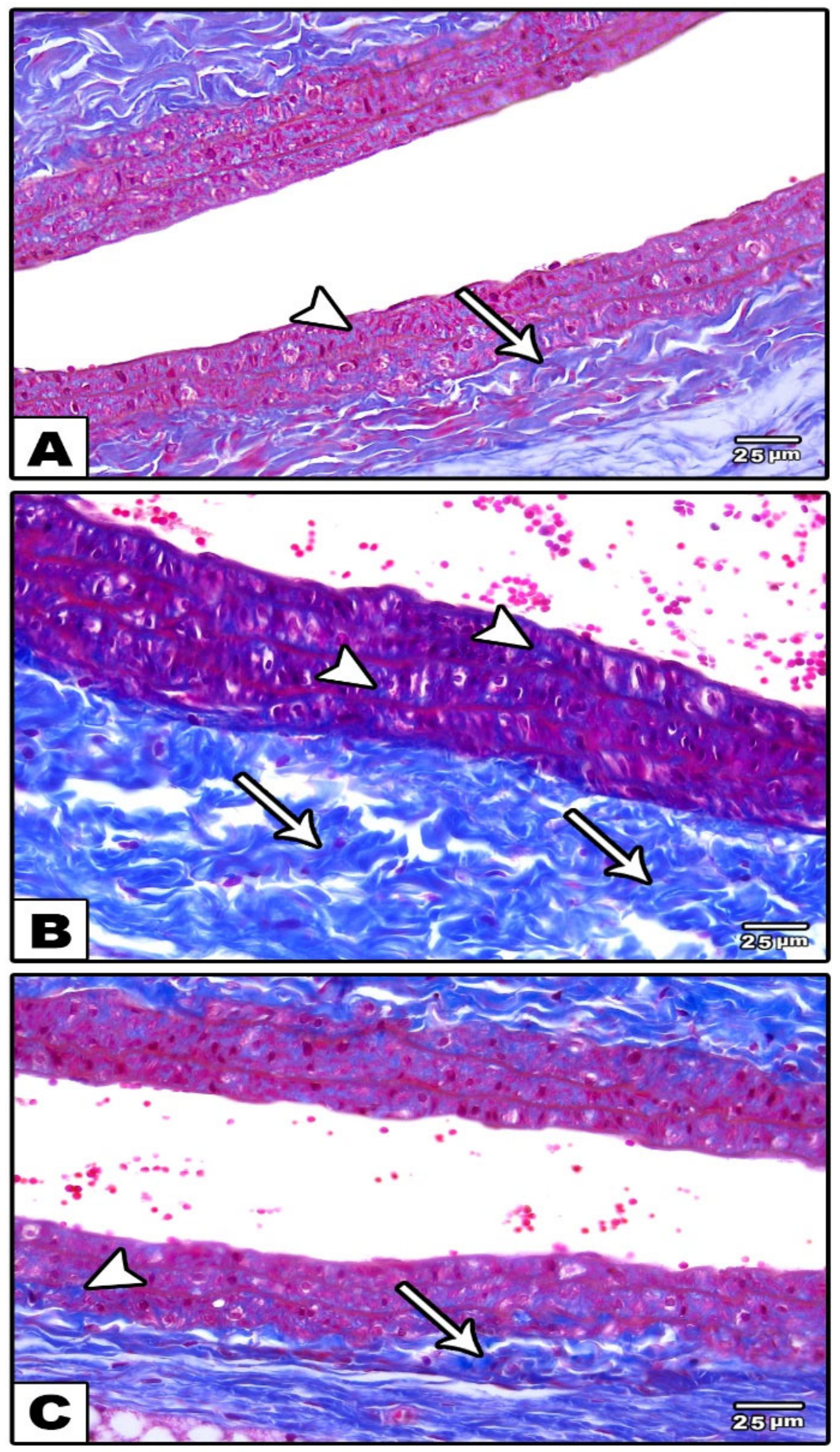
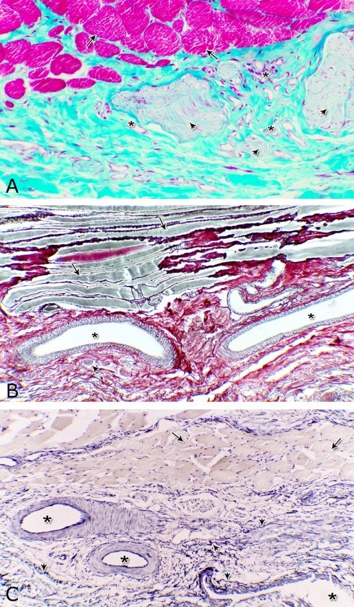
Photomicrograph of Collagen profile Tilapia Skin with and without light ... Download scientific diagram | Photomicrograph of Collagen profile Tilapia Skin with and without light polarization (Picrosirius Red stain, 400x), demonstrating Collagen birefringence yellow-redish ...
PDF Integumentary Answers - Home - Holly H. Nash-Rule, PhD Integumentary Answers - Home - Holly H. Nash-Rule, PhD
Question : Question 31 points Label the photomicrograph of thin skin ... 31 points Label the photomicrograph of thin skin. Hair Follicle Hair Dermis Sebaceous gland Duct of sebaceous gland Reset zoom Solution 5 (1 Ratings ) Solved Biology 2 Years Ago 77 Views This Question has Been Answered! View Solution Question 31) True breeding white flowering plants are crossed to true breeding red flowering plants.



















Post a Comment for "42 photomicrograph of thin skin labeled"